Download
1 / 45
450 likes | 453 Views
AARTI SHARMA M.SC. NURSING OBSTETRICS AND GYNAECOLOGICAL NURSING

E N D
AARTI SHARMA The Urinary System
Functions of the Urinary System • Elimination of waste products • Nitrogenous wastes • Toxins • Drugs Slide 15.1a Copyright © 2003 Pearson Education, Inc. publishing as Benjamin Cummings
Functions of the Urinary System • Regulate aspects of homeostasis • Water balance • Electrolytes • Acid-base balance in the blood • Blood pressure • Red blood cell production • Activation of vitamin D Slide 15.1b Copyright © 2003 Pearson Education, Inc. publishing as Benjamin Cummings
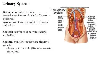
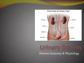
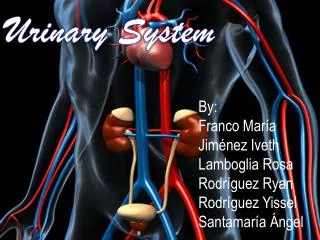
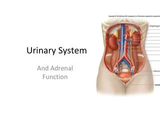
Organs of the Urinary system • Kidneys • Ureters • Urinary bladder • Urethra Figure 15.1a Slide 15.2 Copyright © 2003 Pearson Education, Inc. publishing as Benjamin Cummings
Location of the Kidneys • Against the dorsal body wall • At the level of T12 to L3 • The right kidney is slightly lower than the left • Attached to ureters, renal blood vessels, and nerves at renal hilus • Atop each kidney is an adrenal gland Slide 15.3 Copyright © 2003 Pearson Education, Inc. publishing as Benjamin Cummings
Coverings of the Kidneys • Renal capsule • Surrounds each kidney • Adipose capsule • Surrounds the kidney • Provides protection to the kidney • Helps keep the kidney in its correct location Slide 15.4 Copyright © 2003 Pearson Education, Inc. publishing as Benjamin Cummings
Regions of the Kidney • Renal cortex – outer region • Renal medulla – inside the cortex • Renal pelvis – inner collecting tube Figure 15.2b Slide 15.5 Copyright © 2003 Pearson Education, Inc. publishing as Benjamin Cummings
Kidney Structures • Medullary pyramids – triangular regions of tissue in the medulla • Renal columns – extensions of cortex-like material inward • Calyces – cup-shaped structures that funnel urine towards the renal pelvis Slide 15.6 Copyright © 2003 Pearson Education, Inc. publishing as Benjamin Cummings
Blood Flow in the Kidneys Figure 15.2c Slide 15.7 Copyright © 2003 Pearson Education, Inc. publishing as Benjamin Cummings
Nephrons • The structural and functional units of the kidneys • Responsible for forming urine • Main structures of the nephrons • Glomerulus • Renal tubule Slide 15.8 Copyright © 2003 Pearson Education, Inc. publishing as Benjamin Cummings
Glomerulus • A specialized capillary bed • Attached to arterioles on both sides (maintains high pressure) • Large afferent arteriole • Narrow efferent arteriole Figure 15.3c Slide 15.9a Copyright © 2003 Pearson Education, Inc. publishing as Benjamin Cummings
Glomerulus • The glomerulus sits within a glomerular capsule (the first part of the renal tubule) Figure 15.3c Slide 15.9b Copyright © 2003 Pearson Education, Inc. publishing as Benjamin Cummings
Renal Tubule • Glomerular (Bowman’s) capsule • Proximal convoluted tubule • Loop of Henle • Distal convoluted tubule Slide 15.10 Figure 15.3b Copyright © 2003 Pearson Education, Inc. publishing as Benjamin Cummings
Types of Nephrons • Cortical nephrons • Located entirely in the cortex • Includes most nephrons Figure 15.3a Slide 15.11a Copyright © 2003 Pearson Education, Inc. publishing as Benjamin Cummings
Types of Nephrons • Juxtamedullary nephrons • Found at the boundary of the cortex and medulla Figure 15.3a Slide 15.11b Copyright © 2003 Pearson Education, Inc. publishing as Benjamin Cummings
Peritubular Capillaries • Arise from efferent arteriole of the glomerulus • Normal, low pressure capillaries • Attached to a venule • Cling close to the renal tubule • Reabsorb (reclaim) some substances from collecting tubes Slide 15.12 Copyright © 2003 Pearson Education, Inc. publishing as Benjamin Cummings
Urine Formation Processes • Filtration • Reabsorption • Secretion Figure 15.4 Slide 15.13 Copyright © 2003 Pearson Education, Inc. publishing as Benjamin Cummings
Filtration • Nonselective passive process • Water and solutes smaller than proteins are forced through capillary walls • Blood cells cannot pass out to the capillaries • Filtrate is collected in the glomerular capsule and leaves via the renal tubule Slide 15.14 Copyright © 2003 Pearson Education, Inc. publishing as Benjamin Cummings
Reabsorption • The peritubular capillaries reabsorb several materials • Some water • Glucose • Amino acids • Ions • Some reabsorption is passive, most is active • Most reabsorption occurs in the proximal convoluted tubule Slide 15.15 Copyright © 2003 Pearson Education, Inc. publishing as Benjamin Cummings
Materials Not Reabsorbed • Nitrogenous waste products • Urea • Uric acid • Creatinine • Excess water Slide 15.16 Copyright © 2003 Pearson Education, Inc. publishing as Benjamin Cummings
Secretion – Reabsorption in Reverse • Some materials move from the peritubular capillaries into the renal tubules • Hydrogen and potassium ions • Creatinine • Materials left in the renal tubule move toward the ureter Slide 15.17 Copyright © 2003 Pearson Education, Inc. publishing as Benjamin Cummings
Formation of Urine Figure 15.5 Slide 15.18 Copyright © 2003 Pearson Education, Inc. publishing as Benjamin Cummings
Ureters • Slender tubes attaching the kidney to the bladder • Continuous with the renal pelvis • Enter the posterior aspect of the bladder • Runs behind the peritoneum • Peristalsis aids gravity in urine transport Slide 15.20 Copyright © 2003 Pearson Education, Inc. publishing as Benjamin Cummings
Urinary Bladder • Smooth, collapsible, muscular sac • Temporarily stores urine Figure 15.6 Slide 15.21a Copyright © 2003 Pearson Education, Inc. publishing as Benjamin Cummings
Urinary Bladder • Trigone – three openings • Two from the ureters • One to the urethrea Figure 15.6 Slide 15.21b Copyright © 2003 Pearson Education, Inc. publishing as Benjamin Cummings
Urinary Bladder Wall • Three layers of smooth muscle (detrusor muscle) • Mucosa made of transitional epithelium • Walls are thick and folded in an empty bladder • Bladder can expand significantly without increasing internal pressure Slide 15.22 Copyright © 2003 Pearson Education, Inc. publishing as Benjamin Cummings
Urethra • Thin-walled tube that carries urine from the bladder to the outside of the body by peristalsis • Release of urine is controlled by two sphincters • Internal urethral sphincter (involuntary) • External urethral sphincter (voluntary) Slide 15.23 Copyright © 2003 Pearson Education, Inc. publishing as Benjamin Cummings
Urethra Gender Differences • Length • Females – 3–4 cm (1 inch) • Males – 20 cm (8 inches) • Location • Females – along wall of the vagina • Males – through the prostate and penis Slide 15.24a Copyright © 2003 Pearson Education, Inc. publishing as Benjamin Cummings
Urethra Gender Differences • Function • Females – only carries urine • Males – carries urine and is a passageway for sperm cells Slide 15.24b Copyright © 2003 Pearson Education, Inc. publishing as Benjamin Cummings
Micturition (Voiding) • Both sphincter muscles must open to allow voiding • The internal urethral sphincter is relaxed after stretching of the bladder • Activation is from an impulse sent to the spinal cord and then back via the pelvic splanchnic nerves • The external urethral sphincter must be voluntarily relaxed Slide 15.25 Copyright © 2003 Pearson Education, Inc. publishing as Benjamin Cummings
Maintaining Water Balance • Normal amount of water in the human body • Young adult females – 50% • Young adult males – 60% • Babies – 75% • Old age – 45% • Water is necessary for many body functions and levels must be maintained Slide 15.26 Copyright © 2003 Pearson Education, Inc. publishing as Benjamin Cummings
Distribution of Body Fluid • Intracellular fluid (inside cells) • Extracellular fluid (outside cells) • Interstitial fluid • Blood plasma Figure 15.7 Slide 15.27 Copyright © 2003 Pearson Education, Inc. publishing as Benjamin Cummings
The Link Between Water and Salt • Changes in electrolyte balance causes water to move from one compartment to another • Alters blood volume and blood pressure • Can impair the activity of cells Slide 15.28 Copyright © 2003 Pearson Education, Inc. publishing as Benjamin Cummings
Maintaining Water Balance • Water intake must equal water output • Sources for water intake • Ingested foods and fluids • Water produced from metabolic processes • Sources for water output • Vaporization out of the lungs • Lost in perspiration • Leaves the body in the feces • Urine production Slide 15.29 Copyright © 2003 Pearson Education, Inc. publishing as Benjamin Cummings
Maintaining Water Balance • Dilute urine is produced if water intake is excessive • Less urine (concentrated) is produced if large amounts of water are lost • Proper concentrations of various electrolytes must be present Slide 15.30 Copyright © 2003 Pearson Education, Inc. publishing as Benjamin Cummings
Regulation of Water and Electrolyte Reabsorption • Regulation is primarily by hormones • Antidiuretic hormone (ADH) prevents excessive water loss in urine • Aldosterone regulates sodium ion content of extracellular fluid • Triggered by the rennin-angiotensin mechanism • Cells in the kidneys and hypothalamus are active monitors Slide 15.31 Copyright © 2003 Pearson Education, Inc. publishing as Benjamin Cummings
Maintaining Acid-Base Balance in Blood • Blood pH must remain between 7.35 and 7.45 to maintain homeostasis • Alkalosis – pH above 7.45 • Acidosis – pH below 7.35 • Most ions originate as byproducts of cellular metabolism Slide 15.33a Copyright © 2003 Pearson Education, Inc. publishing as Benjamin Cummings
Maintaining Acid-Base Balance in Blood • Most acid-base balance is maintained by the kidneys • Other acid-base controlling systems • Blood buffers • Respiration Slide 15.33b Copyright © 2003 Pearson Education, Inc. publishing as Benjamin Cummings
Blood Buffers • Molecules react to prevent dramatic changes in hydrogen ion (H+) concentrations • Bind to H+ when pH drops • Release H+ when pH rises • Three major chemical buffer systems • Bicarbonate buffer system • Phosphate buffer system • Protein buffer system Slide 15.34 Copyright © 2003 Pearson Education, Inc. publishing as Benjamin Cummings
The Bicarbonate Buffer System • Mixture of carbonic acid (H2CO3) and sodium bicarbonate (NaHCO3) • Bicarbonate ions (HCO3–) react with strong acids to change them to weak acids • Carbonic acid dissociates in the presence of a strong base to form a weak base and water Slide 15.35 Copyright © 2003 Pearson Education, Inc. publishing as Benjamin Cummings
Respiratory System Controls of Acid-Base Balance • Carbon dioxide in the blood is converted to bicarbonate ion and transported in the plasma • Increases in hydrogen ion concentration produces more carbonic acid • Excess hydrogen ion can be blown off with the release of carbon dioxide from the lungs • Respiratory rate can rise and fall depending on changing blood pH Slide 15.36 Copyright © 2003 Pearson Education, Inc. publishing as Benjamin Cummings
Renal Mechanisms of Acid-Base Balance • Excrete bicarbonate ions if needed • Conserve or generate new bicarbonate ions if needed • Urine pH varies from 4.5 to 8.0 Slide 15.37 Copyright © 2003 Pearson Education, Inc. publishing as Benjamin Cummings
Developmental Aspects of the Urinary System • Functional kidneys are developed by the third month • Urinary system of a newborn • Bladder is small • Urine cannot be concentrated Slide 15.38a Copyright © 2003 Pearson Education, Inc. publishing as Benjamin Cummings
Developmental Aspects of the Urinary System • Control of the voluntary urethral sphincter does not start until age 18 months • Urinary infections are the only common problems before old age Slide 15.38b Copyright © 2003 Pearson Education, Inc. publishing as Benjamin Cummings
Aging and the Urinary System • There is a progressive decline in urinary function • The bladder shrinks with aging • Urinary retention is common in males Slide 15.39 Copyright © 2003 Pearson Education, Inc. publishing as Benjamin Cummings