Download

1 / 43
0 likes | 7 Views
Join me for 'Plant Anatomy Unveiled' - a brief journey into the hidden world of plant tissues. Delve into the intricate structures of dicot and monocot tissues, witness the wonders of vascular bundles, and uncover the remarkable adaptations that fuel plant resilience. This presentation is your passport to a visual odyssey, offering a condensed yet immersive experience into the fascinating realm of plant anatomy.

E N D
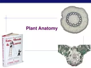
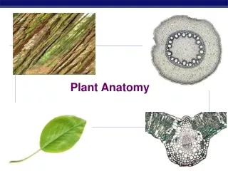
Plant Anatomy PPT模板http://www.1ppt.com/moban/
CONTENTS 01 02 01 Vascular Bundles Anatomy of Dicot Root 02 04 03 Anatomy of Monocot Root Anatomy of Dicot Stem 04 05 Anatomy of Monocot Stem 05
01 Vascular Bundles
Vascular Bundles • A vascular bundle is a crucial structural component found in the stems and roots of plants. It serves as the plant's transport system, responsible for the movement of water, nutrients, and sugars throughout the plant. Vascular bundles consist of two primary tissues: xylem, which transports water and minerals from the roots to the rest of the plant, and phloem, which carries sugars and other organic compounds from the leaves to various plant parts. In dicotyledonous plants, the vascular bundle often includes a cambium layer, which enables secondary growth, increasing the plant's girth over time. Vascular bundles come in various types, including collateral, concentric, radial, stellate, and cortical, depending on their arrangement and composition, reflecting the plant's specific needs and adaptations. These bundles are essential for plant growth, providing support and facilitating the vital processes of nutrient and water transport.
Xylem • Xylem is one of the two main vascular tissues in plants, the other being phloem. It is a vital plant tissue responsible for the transport of water, minerals, and other dissolved substances from the roots to the rest of the plant. It also provides structural support to the plant. Here's a comprehensive overview of xylem, including its definitions, types, anatomy, and exceptions:
Types of Xylem Primary Xylem: This is formed during the primary growth of a plant and is present in the young parts of stems and roots. Secondary Xylem: Also known as wood, secondary xylem is formed during secondary growth and is responsible for the thickening of stems and roots. It is the type of xylem that most people associate with trees.
Anatomy of Xylem: Xylem consists of several types of specialized cells, each with a specific role in water transport and support. Tracheids Vessel elements These are long, tapering cells with thick lignified walls. They are found in both primary and secondary xylem and serve as conduits for water transport. Tracheids are common in gymnosperms and some angiosperms. Vessel elements are wider and shorter than tracheids and are found in the xylem of most angiosperms (flowering plants). They have perforated end walls called perforation plates, which facilitate efficient water flow. Xylem Parenchyma Xylem Fibres These are living cells found in between tracheids and vessel elements. They store starch and provide structural support to the xylem. Fibre cells have thick walls and provide additional structural support to the xylem.
Exceptions in Xylem • Gymnosperms vs. Angiosperms: • Gymnosperms, such as pine trees, primarily have tracheids in their xylem, while angiosperms, including most flowering plants, have both tracheids and vessel elements. • Vesselless Angiosperms: • Some primitive angiosperms, like the water lily family (Nymphaeaceae) and Amborella, lack vessel elements in their xylem, relying solely on tracheids for water transport. • Lack of Secondary Xylem: • Some herbaceous plants or annuals may not develop secondary xylem because they don't undergo significant secondary growth. Their xylem remains in a primary state. • Xylem is a remarkable tissue that plays a crucial role in maintaining the plant's water balance, enabling it to thrive in terrestrial environments. Its diverse cell types and adaptations demonstrate the remarkable diversity of plants and their ability to adapt to various ecological niches.
Functions of Xylem • Water Transport: • The primary function of xylem is to transport water and dissolved minerals from the roots to the rest of the plant. This process is essential for maintaining the plant's hydration, allowing it to carry out various physiological processes like photosynthesis, respiration, and transpiration. • Mineral Transport: • Along with water, xylem also carries essential minerals and nutrients absorbed from the soil. These minerals, including potassium, calcium, and magnesium, are crucial for plant growth, enzyme activation, and various metabolic processes. • Support: • Xylem provides structural support to the plant. The lignified cell walls of tracheids, vessel elements, and fiber cells contribute to the plant's rigidity and help it withstand the forces of gravity and wind.
Functions of Xylem • Transpiration Pull: • Xylem plays a key role in the transpiration process, where water evaporates from the leaves through tiny openings called stomata. This loss of water creates a negative pressure gradient, known as the transpiration pull, which draws water up through the xylem from the roots to the leaves. • Storage: • Parenchyma cells in the xylem can store starch and other carbohydrates, which can be mobilized when the plant needs energy or nutrients during periods of growth or stress. • Defense: • In some cases, xylem cells can help defend the plant against pathogens and pests. For example, certain phenolic compounds and resins produced by xylem cells can inhibit the growth of microorganisms and deter herbivores. • In summary, xylem is a critical tissue in plants that serves multiple vital functions. It facilitates the transport of water, minerals, and nutrients, provides structural support to the plant, generates the transpiration pull necessary for water uptake, and even contributes to defense mechanisms in certain plant species. Without xylem, plants would be unable to thrive in terrestrial environments and perform essential life processes.
Phloem • Phloem is a complex plant tissue responsible for the transport of organic nutrients, primarily sugars (mainly sucrose), from the leaves (source) to various parts of the plant, including roots, stems, and developing fruits and seeds (sink).
Elements of Phloem Sieve Tube Elements: These are the main cells responsible for sugar transport. They are long, slender cells with perforated end walls called sieve plates, through which sugars flow. Sieve tube elements are alive but lack a nucleus and other organelles at maturity. Companion Cells: These are small, nucleated cells closely associated with sieve tube elements. Companion cells provide metabolic support to sieve tube elements, helping maintain their function. Phloem Parenchyma: Similar to xylem, the phloem also contains parenchyma cells that store and transport various nutrients.
Exceptions in Phloem • Lack of Sieve Tubes: • In certain non-vascular plants like mosses and liverworts, as well as in some primitive vascular plants like ferns, the phloem lacks true sieve tubes. Instead, these plants have simpler food-conducting cells. • Lack of Companion Cells: • Some gymnosperms, such as conifers, have sieve tube elements that lack companion cells. Instead, specialized parenchyma cells fulfill some of the companion cell functions. • Occurrence of Extra-Phloem Tissues: • In some cases, non-phloem tissues can also be involved in sugar transport. Fr example, in certain monocotyledonous plants, the transport of sugars occurs through extra-phloem tissues known as "vascular bundles with internal phloem. • Phloem is essential for the distribution of nutrients and energy within a plant, ensuring its growth, development, and survival. Its complexity and ability to transport various substances make it a critical component of the plant's vascular system.
Functions of Phloem • Sugar Transport: • The primary function of phloem is the translocation of sugars produced during photosynthesis in the leaves to other parts of the plant for growth, energy storage, and various metabolic processes. • Nutrient Transport: • Phloem can also transport other organic compounds, including amino acids and hormones, to different plant organs. • Bidirectional Flow: • Phloem allows bidirectional flow, meaning it can transport sugars not only from leaves to sinks but also in the reverse direction when needed (e.g., during the winter to support root growth).
Cambium • The cambium is a vital tissue found in the vascular bundles of plants, primarily in woody plants, and is crucial for their growth and development. It is a thin layer of actively dividing cells located between the xylem and phloem tissues in the plant stem and roots. The cambium plays a fundamental role in secondary growth, which is responsible for increasing the girth or thickness of the plant over time, leading to the development of woody tissues.
Key Points about Cambium • Cell Division: • The cambium consists of meristematic cells that undergo rapid cell division. These cells divide to produce new cells, which can differentiate into xylem cells towards the inside and phloem cells towards the outside. • Secondary Growth: • The primary function of the cambium is to contribute to secondary growth, which is responsible for the increase in diameter or girth of the plant. Secondary growth allows plants to become taller and thicker over time, making them • suitable for long-term growth and structural support. • Xylem and Phloem Formation: • As the cambium cells divide and differentiate, they give rise to xylem cells on the inner side and phloem cells on the outer side. Xylem cells are responsible for transporting water and minerals from the roots to the rest of the plant, while phloem cells transport nutrients produced during photosynthesis from the leaves to other parts of the plant. • Annual Growth Rings: • In many woody plants, the activity of the cambium varies with the seasons, resulting in the formation of annual growth rings. These rings can be counted to determine the age of the plant and provide information about its environmental conditions during different years.
Key Points about Cambium • Cambial Activity: • Cambial activity is influenced by various factors, including hormonal signals, environmental conditions, and the plant's physiological state. Changes in cambial activity can result in variations in wood density, which can be important in the timber industry. • In summary, the cambium is a critical tissue in vascular bundles that facilitates secondary growth in plants. It is responsible for the formation of new xylem and phloem cells, contributing to the plant's increasing thickness and enabling it to adapt and thrive in various environmental conditions.
Types of Vascular Bundles Radial Vascular Bundle • A radial vascular bundle is a type of plant vascular tissue arrangement commonly found in the roots of angiosperms. In this vascular bundle, xylem and phloem tissues are found radially, alternating with each other, and equal in number. This is the most primitive type of vascular bundle and helps to provide structural support. Conjoint Vascular Bundle • Conjoint vascular bundles are plant structures that contain both xylem and phloem tissues arranged together for the efficient transport of water, nutrients, and sugar. There are three main types of conjoint vascular bundles: collateral, bicollateral, and concentric.
Conjoint vascular bundles are further categorized as: • Collateral Vascular Bundle: • In a collateral vascular bundle, xylem and phloem tissues are positioned adjacent to each other within the bundle where the xylem tissue is usually located toward the center, while the phloem tissue surrounds it. • In dicot stem, a cambium strip is present in these bundles between xylem and phloem tissues, unlike monocot stem. The former is called open collateral vascular bundle whereas, the latter is called closed collateral vascular bundle. • Bicollateral Vascular Bundle: • Bicollateral vascular bundles are similar to collateral bundles but have an additional layer of phloem tissue on both the inner and outer sides of the xylem. • This type of bundle is often found in stem of members of Solanaceae, Cucurbitaceae and Convolvulaceae. • Concentric Vascular Bundle: • Concentric vascular bundles have a different arrangement, where one of the two vascular tissues (xylem & phloem) forms the central core and the other surrounds it completely on all sides. Concentric vascular bundles are found in rhizome of fern plants where xylem forms the central core. They are also found in stem of Dracaena, Yucca etc. where phloem forms the central core.
02 Anatomy of Dicot Root
Anatomy of Dicot Root • To understand the inner workings of a dicot root, it's essential to delve into its intricate anatomy and the specialized structures it harbors. • In the following sections, we'll explore the various components that make up a dicot root, each with its unique role in supporting the plant's growth and function.
Anatomy of Dicot Root • Epiblema : • The epiblema, also known as the root cap, is a protective structure at the root tip. Composed of parenchyma cells, it serves as a shield, preventing damage to the delicate growing tip. The epiblema secretes mucilage, aiding in soil penetration, and absorption of water and mineral salts from soil. Additionally, it contains statocytes that sense gravity, helping the root grow in the correct direction. • Cortex: • The cortex is a critical region located just beneath the epiblema. It primarily consists of parenchyma cells with sufficient intercellular spaces among them, which store food reserves and provide mechanical support to the root. The cortex also plays a vital role in water conduction from epiblema to inner tissues. • Endodermis: • The endodermis is a single layer of specialized barrel-shaped cells surrounding the vascular cylinder. It has a prominent role in regulating the movement of water and nutrients into the vascular tissues. • The radial walls of endodermal cells are impregnated with Suberin, a waxy substance, forming the Casparian strip, which acts as a selective barrier. • Pericycle: • The pericycle is a layer of parenchymatous cells located just inside the endodermis. It is responsible for giving rise to lateral roots during secondary growth in dicot roots. In some cases, the pericycle can also undergo meristematic activity, contributing to root thickening.
Anatomy of Dicot Root • Vascular Bundles: • Dicot roots typically exhibit a radial arrangement of vascular bundles. Each vascular bundle consists of alternating radii of xylem (towards the center) and phloem (towards the periphery). The number of vascular bundles in dicot roots can vary, but it typically ranges from 2 to 6. • Xylem: • Xylem tissue in dicot roots transports water and minerals absorbed from the soil to the rest of the plant. It is composed of various cell types, including tracheids, vessel elements, xylem parenchyma and xylem fibres. Xylem cells are responsible for the upward movement of water and nutrients. • Phloem: • Phloem tissue carries sugars and other organic compounds produced in the leaves to different parts of the plant. It includes sieve tube elements and companion cells, which work together to facilitate efficient nutrient transport. Phloem is responsible for the downward movement of sugars within the plant.
Anatomy of Dicot Root • Conjunctive Tissue: • Conjunctive tissue is a non-vascular tissue consisting of compactly arranged parenchyma cells, found in between xylem and phloem bundles. It provides structural support to the root and also plays a role in nutrient storage. • Pith: • The pith is the central, often parenchymatous, tissue within the vascular cylinder of a dicot root. It can serve as a storage region for nutrients and provides additional structural support to the root.
03 Anatomy of Monocot Root
Anatomy of Monocot Root • Understanding the anatomy of a monocot root is essential for comprehending the unique structural and functional adaptations that monocots have developed to meet their specific growth and nutrient transport needs. • Let's examine the complex architecture and specialized structures of a monocot root to better comprehend how it functions.
Anatomy of Monocot Root • Epiblema: • The epiblema, or root cap, is the outermost single-layered protective structure of a monocot root. • Composed of parenchyma cells, its primary function is to shield the delicate root tip from abrasion and facilitate soil penetration. Velamen is a specialized layer in the epiblema of some monocot roots, made up of multiple layers of dead, spongy cells. They are found in epiphytic monocots such as Orchids and take part in moisture absorption from atmosphere. • Cortex: • The cortex is a multi-layered, wide parenchymatous region found just beneath the epidermis; which stores food reserves, conducts water and minerals upto endodermis, and may contain air spaces for aeration. In old monocot roots, a few layers (or single layer) of cortex just beneath the epidermis give rise to a thick-walled sclerenchymatous region called exodermis. This layer provides mechanical support and protects internal tissues from injurious agencies. • Endodermis: • The endodermis in a monocot root is a single layer of tightly packed cells surrounding the vascular cylinder. Similar to dicot roots, the endodermal cells in monocots may have a Casparian strip, a waxy barrier. So, endodermis plays a crucial role in controlling the movement of water and nutrients into the vascular tissues. Thus, it is also known as “biological check-post”.
Anatomy of Monocot Root • Pericycle: • The pericycle is a layer of cells located just inside the endodermis, though it is often less prominent in monocot roots compared to dicots. It can give rise to lateral roots but is not involved in secondary growth as in dicot roots. • Vascular Bundles: • Monocot roots have a different vascular bundle arrangement compared to dicots. They typically exhibit a scattered or atactostele arrangement, where individual vascular bundles are spread throughout the root. The vascular bundles in monocot roots lack a clearly defined arrangement of xylem and phloem radii, as seen in dicots. • Monocot roots have a radial arrangement of vascular bundles in which xylem and phloem tissues are found alternating with each other at different radii. • Their number can vary from 8 to 20, but it may reach upto 100 in case of Pandanus. • Xylem: • In monocot roots, the xylem tissue is typically found toward the center of each vascular bundle. It transports water and minerals from the root to the rest of the plant and consists of various cell types, including vessel elements and tracheids. • Phloem: • Phloem tissue in monocot roots is usually located toward the periphery of each vascular bundle. It transports sugars and other organic compounds produced in the leaves to different plant parts. Monocot phloem typically contains sieve tube elements and companion cells with a few phloem parenchyma.
Anatomy of Monocot Root • Conjunctive Tissue: • Conjunctive tissue is the non-vascular tissue that surrounds and supports the vascular bundles in monocot roots. • It helps maintain the integrity and organization of the root structure. • Pith: • It is a wide parenchymatous zone found at the centre of the root. The pith stores food materials, and thus the pith cells contain abundant amount of starch grains.
Differences between Dicot & Monocot Root • Monocot Root • Casparian strips are visible only in young root. • Epiblema is mostly single layered but it is found in multilayered form in some epiphytic plants like Orchids. • Pericycle maybe parenchymatous or sclerenchymatous. • Secondary growth is absent. • Pith is well-developed. • Xylem and phloem bundles are usually 8 to 20 in number. Dicot Root • Endodermis is less thickened and Casparian strips are more prominent. • Epiblema is always single layered. • Pericycle is parenchymatous • Secondary growth takes place with the help of vascular cambium and cork cambium. • Pith is either absent or greatly reduced. • Number of xylem and phloem bundles varies from 2 to 6 but sometimes 8.
04 Anatomy of Dicot Stem
Anatomy of Dicot Stem • Understanding the anatomy of a dicot stem is fundamental for gaining insight into the distinctive structural and functional adaptations that dicotyledonous plants have evolved to cater to their specific requirements for growth, structural support, and the efficient transport. • Exploring the intricate design and unique components of a dicot stem will enhance our understanding of its operational mechanisms.
Anatomy of Dicot Stem • Epidermis: • The epidermis is the outermost layer of a dicot stem. Comprising a single layer of tightly packed cells, it serves as a protective barrier against environmental stressors. The epidermis often contains stomata, which regulate gas exchange whereas the presence of cuticle layer on epidermis helps to reduce rate of transpiration. • Hypodermis: • Located just below the epidermis, the hypodermis is a zone of multiple layers of collenchyma. The cells bear chloroplast and take part in photosynthesis. This region provides mechanical support and elasticity to the stem. • General Cortex: • The general cortex of a dicot stem serves as a versatile region that supports the plant's growth, storage, gas exchange, and healing processes. Its parenchymatous cells, with their flexible nature and ability to store reserves, contribute to the stem's overall health and functionality. • Endodermis: • The stem endodermis consists of a single layer of tightly packed cells. It lacks Casparian strips, thus it is also known as “endodermoid”. Cells of endodermis contain large amount of starch grain. Endodermis takes part in selective absorption, and water and nutrient regulation.
Anatomy of Dicot Stem • Pericycle: • The pericycle in a dicot stem is a versatile layer of cells with critical roles in lateral root initiation, secondary growth in some dicot species, adaptation to changing environmental conditions, and enhancing nutrient absorption. Its ability to generate new roots and contribute to stem thickness is essential for the plant's development and survival in diverse habitats. • Vascular Bundles: • In dicot stems, vascular bundles are typically arranged in a circular pattern or a ring around central pith. This type of arrangement of vascular bundles is called “Eustele”. The vascular bundles in dicot stem are conjoint, collateral, open and wedge-shaped. • Xylem: • Xylem tissue in dicot stems facilitates the upward transport of water and minerals from the roots to the rest of the plant. It includes tracheids, vessels, xylem parenchyma and xylem fibres; characterized by lignified cell walls. • Cambium: • A strip of cambium is found in between xylem and phloem tissues. It is also called intrafascicular cambium. It takes part in formation of primary phloem and primary xylem on outer and inner side respectively.
Anatomy of Dicot Stem • Phloem: • Phloem tissue in dicot stems transports organic compounds produced in leaves to different parts of the plant. It comprises sieve tube elements, phloem parenchyma and companion cells that facilitate the efficient transport of nutrients. • Medullary ray: • It is a parenchymatous zone found in between two adjacent vascular bundles. It consists of thin-walled cells that take part in food storage and material conduction from pith to cortex and vice versa. • Pith: • The pith is the central, parenchymatous tissue within the stem. It stores nutrients and may add internal support to the stem.
05 Anatomy of Monocot Stem
Anatomy of Monocot Stem • Comprehending the anatomy of a monocot stem is pivotal in uncovering the specialized structural and functional adaptations that monocotyledonous plants have evolved to address their specific growth, structural, and transport necessities. • Exploring the intricate arrangement and distinctive elements of a monocot stem will enrich our insight into its operational dynamics.
Anatomy of Monocot Stem • Epidermis: • The outermost layer of the stem, the epidermis is a single layer of tightly packed cells that protects the stem. It is covered by a thick cuticle, which helps to prevent rate of transpiration. The epidermis also contains stomata, which are openings that allow gas exchange. • Hypodermis: • The hypodermis is a layer of cells that lies just beneath the epidermis. The cells of the hypodermis are typically sclerenchyma cells, which are thick-walled and lignified and provide support for the stem. • Ground Tissue: • In monocot stem, the differentiation of cortex, endodermis, pericycle, medullary rays and pith does not take place but a wide parenchyma zone is found from hypodermis to the center. It is called the ground tissue. The ground tissue is the bulk of the stem. It has a variety of functions, including photosynthesis, storage, and transport. The ground tissue also contains the vascular bundles.
Anatomy of Monocot Stem • Vascular Bundles: • Monocot stems typically exhibit a scattered arrangement of vascular bundles which is known as “Atactostele” arrangement. Each vascular bundle consists of xylem and phloem tissues. The xylem and phloem tissues are surrounded by a sheath of sclerenchyma cells called the bundle sheath. • Xylem: • Xylem tissue in monocot stems is centrally located within each vascular bundle. It transports water and minerals from the roots to various parts of the plant. Monocot xylem consists of vessel elements and tracheids, both characterized by lignified cell walls. • Phloem: • Phloem tissue is situated at the periphery of each vascular bundle in monocot stems. Its main function is to transport sugars and other organic compounds produced in the leaves to different plant regions. Monocot phloem contains sieve tube elements and companion cells, which aid in nutrient transport.
Differences between Dicot & Monocot Root • Monocot Stem • Endodermis and pericycle are not differentiated. • Vascular bundles are surrounded by sclerenchymatous bundle sheath. • Hypodermis is sclerenchymatous. • Vascular bundles are scattered. • Pith and medullary rays are not differentiated. • Vascular bundles are closed collateral type and oval-shaped. Dicot Stem • Endodermis and pericycle are well-differentiated. • Vascular bundles are not covered. • Hypodermis is usually collenchymatous. • Vascular bundles are arranged in a ring. • Pith and medullary rays are clearly distinct. • Vascular bundles are open collateral type and wedge-shaped.
Presented by: Shishir Pokharel, 40 XII CMilan Khatri, 22 XII C
Any Queries? E-mail me at: sisirpokharel27@gmail.com