Download
1 / 68
690 likes | 697 Views
Educative presentation for medical graduates and postgraduates

E N D
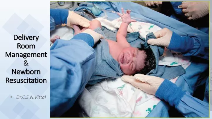
Delivery Room Management&Newborn Resuscitation Dr.C.S.N.Vittal
Principles • Birth asphyxia accounts for about 1/4th of neonatal deaths occur each year worldwide • 90% of newborns make smooth transition from intrauterine to extrauterine life requiring little assistance • 10% of newborns need some assistance • Only 1% require extensive resuscitation
How does a baby receive oxygen before birth? Fetal Circulation Transitional Circulation
How does a baby receive oxygen before birth? • All the oxygen used by a fetus diffuses across the placental membrane from the mother’s blood to the baby’s blood. • Only a small fraction of fetal blood passes through the fetal lungs. • Most of the blood from the right side of the heart cannot enter the lungs because of the increased resistance to flow in the constricted blood vessels in the fetal lungs
How does a baby receive oxygen before birth? • Most of this blood takes the lower resistance path through the ductus arteriosus into the aorta
What happens normally at birth? • fluid in the alveoli is absorbed into pulmonary lymphatics and replaced by air • Umbilical arteries constrict and then the umbilical arteries and vein are closed when the umbilical cord is clamped. • Blood vessels in the lung tissue relax, decreasing resistance to blood flow
What can go wrong? • Compromise of uterine or placental blood flow > deceleration of FHR • Weak cry > inadequate ventilation to push alveolar fluid • In utero hypoxia > Meconium passage > may block airways • Fetal blood loss (abruption) > Systemic hypotension • Fetal hypoxia / ischemia > poor cardiac contractility and fetal bradycardia > systemic hypotension • Pulmonary arterioles remain constricted > PPHN
Consequences Compromised baby • Depression of respiratory drive - apnea, tachypnoea • Poor muscle tone • Bradycardia • Low blood pressure • Persistent cyanosis or low SpO2 • These outcomes can be improved with timely and effective resuscitation
How can you tell if a newborn had in uteroor perinatal compromise? • Perinatal stress results in an initial period of rapid breathing followed by a period of primary apnea (no breathing or gasping) • During this period of primary apnea, stimulation, such as drying the newborn or slapping the feet, will cause a resumption of breathing.
How can you tell if a newborn had in uteroor perinatal compromise? • If cardiorespiratory compromise continues during primary apnea, the baby will have an additional brief period of gasping breaths and then will enter a period of secondary apnea • During secondary apnea, stimulation will not restart the baby’s breathing. • Assisted ventilation must be provided to reverse the process.
Sequence of EventsReadiness for neonatal resuscitation requires: 1. Introduction (Preparing) 6. Assessment of need / Administration of O2 2. Anticipation of Resuscitation Need 7. Positive Pressure Ventilation (PPV) 3. Umbilical Cord Management 8. Advanced Assisted Ventilation 9. Chest Compressions 4. Initial Steps 5. Assessment of Heart Rate 10. Medications and Volume Administration
1. IntroductionAnswers to the following 4 pre-birth questions: • Expected gestational age? • Is the AF clear? • Any additional risk factors? • Umbilical cord management plan
Answers to the following 3 questions: • Term gestation? • Good tone? • Breathing or crying? • If the answer to any of the three assessment questions is “No,” the infant should be moved to a radiant warmer to receive one or more of the following 4 actions in sequence: • Initial steps in stabilization (warm and maintain normal temperature, position, clear secretions only if copious and/or obstructing the airway, dry, stimulate) • Ventilate and oxygenate • Initiate chest compressions • Administer epinephrine and/or volume. Approximately 60 seconds (“the Golden Minute”) are allotted for completing the initial steps, re-evaluating, and beginning ventilation if required
2. Anticipation of resuscitation need • Assessment of perinatal risk, • A system to assemble the appropriate personnel based on perinatal risk, • An organized method for ensuring immediate access to supplies and equipment, • Standardization of behavioral skills that help assure effective teamwork and communication
3. Umbilical cord management • Delayed cord clamping for longer than 30 seconds is reasonable for both term and preterm infants who do not require resuscitation at birth. • Routine use of cord milking for infants born at less than 29 weeks of gestation outside of a research setting, NOT RECOMMENDED
4. Initial steps of newborn resuscitation Yes, stay with the mother Birth Routine care • Maintain normal temperature • Position the infant in a “sniffing” position to open the airway • Clear secretions if needed with a bulb syringe or suction catheter • Dry the infant • Ongoing evaluation Term gestation? Breathing or crying? Good tone? 30 sec 60 sec
4. Initial steps of newborn resuscitation Birth Yes, stay with the mother Routine care • Maintain normal temperature • Position the infant in a “sniffing” position to open the airway • Clear secretions if needed with a bulb syringe or suction catheter • Dry the infant • Stimulate Term gestation? Breathing or crying? Good tone? No 30 sec • Clear secretions • Dry the infant • Stimulate 60 sec
4.1 Importance of maintaining normal temperature • Hypothermia is associated with serious morbidities, such as increased risk of intraventricular hemorrhage, respiratory issues, hypoglycemia and late-onset sepsis. • It is recommended that the temperature of newly born nonasphyxiated infants be maintained between 36.5°C and 37.5°C after birth through admission and stabilization. • The goal is an axillary temperature between 36.5°C and 37.5°C.
Interventions to Maintain Newborn Temperature in the Delivery Room • The use of radiant warmers and plastic wrap with a cap • has improved but not eliminated the risk of hypothermia in preterm infants in the delivery room. • To prevent hypothermia in infants born at less than 32 weeks of gestation. • Increased room temperature, • Thermal mattresses, and • Use of warmed humidified resuscitation gases. • All resuscitation procedures, including endotracheal intubation, chest compression, and insertion of intravenous lines, can be performed with these temperature-controlling interventions in place • Hyperthermia (greater than 38.0°C) should be avoided due to the potential associated risks.
Provide warmth Vigorous, term newborn. Initial steps are performed skin-to-skin with mother.
Maintaining Normothermia inResource-Limited Settings • In resource-limited settings, to maintain body temperature or prevent hypothermia during transition (birth until 1 to 2 hours of life) in well newborn infants, it may be reasonable to put them in a clean food- grade plastic bag up to the level of the neck and swaddle them after drying.
Maintaining Normothermia inResource-Limited Settings • Another option that may be reasonable is to nurse such newborns with skin-to-skin contact or kangaroo mother care.
Concept of Warm Chain • Thermal care in delivery room • Delivery room should be 25 -280C • All the linen, cloths should be prewarmed • Radiant warmer should be switched on in advance and put in to manual mode with 100% heater output • Warm resuscitation • Immediate drying • Skin to skin contact • Breast feeding • Postpone bathing/weighing • Clothing and bedding-in • Warm transportation • Training awareness • Polythene occlusive wraps – for preterm • Incubators • KMC
4.2 Clearing the airway When amniotic fluid is clear: • Avoiding unnecessary suctioning helps prevent the risk of induced bradycardia that can be associated with suctioning of the nasopharynx. • It is recommended that suctioning immediately following birth (including suctioning with a bulb syringe) should be reserved for babies who have obvious obstruction to spontaneous breathing or who require positive-pressure ventilation (PPV)
Clearing the airway - When meconium is present: • If the infant born through meconium-stained amniotic fluid presents with poor muscle tone and inadequate breathing efforts, the initial steps of resuscitation should be completed under the radiant warmer. • PPV should be initiated • if the infant is not breathing or • the heart rate is less than 100/min after the initial steps are completed. • Routine intubation for tracheal suction in this setting is not suggested • Emphasis should be made on initiating ventilation within the first minute of life in nonbreathing or ineffectively breathing infants.
5. Assessment of hart rate • The use of 3-lead ECG for the rapid and accurate measurement of the newborn’s heart rate may be reasonable. • Pulse oximeter tended to underestimate the newborn’s heart rate (potentially leading to unnecessary interventions)
5. Assessment of hart rate Alternative methods for assessing the heart rate: pulse oximetry and ECG monitor
6. Assessment of oxygen need and adm • Clinical assessment of skin color is a very poor indicator of oxyhemoglobin saturation during the immediate neonatal period and lack of cyanosis appears to be a very poor indicator of the state of oxygenation of an uncompromised baby following birth. • There is evidence that either insufficient or excessive oxygenation can be harmful to the newborn infant.
6.1 Use of Pulse Oximetry • It is recommended that oximetry be used when resuscitation can be anticipated, when PPV is administered, when central cyanosis persists beyond the first 5 to 10 minutes of life, or when supplementary oxygen is administered. • The probe should be attached to a preductal location (ie, the right upper extremity, usually the wrist or medial surface of the palm).
6.2 Administration of oxygen • In term and late-preterm newborns (≥35 weeks of gestation) receiving respiratory support at birth, the initial use of 21% oxygen is reasonable. • One hundred percent oxygen should not be used to initiate resuscitation because it is associated with excess mortality. • Oxygen concentration should be titrated to achieve a preductal oxygen saturation approximating the interquartile range (see table) measured in healthy term babies following vaginal birth at sea level.
6.2 Administration of oxygen • In preterm newborns (<35 weeks of gestation) receiving respiratory support at birth, it may be reasonable to begin with 21% to 30% oxygen with subsequent oxygen titration based on pulse oximetry. • Oxygen concentration should be titrated to achieve a preductal oxygen saturation approximating the interquartile range (see table) measured in healthy term babies following vaginal birth at sea level.
7. Positive Pressure Ventilation (PPV) • If the infant remains apneic or gasping, or • If the heart rate remains <100 per minute after performing the initial steps, begin positive pressure ventilation.
Different Types of Resuscitation Devices Flow inflating bags Fills only when oxygen from a compressed source flows in to it Self inflating bags Fills spontaneously after it is squeezed, pulling oxygen or air in to the bag T-piece resuscitator Also works when gas from compressed source flows into it. The gas is directed into the baby by occluding the opening on T-piece
T-Piece Resuscitator Directs compressed gas toward the baby when an opening on the top of the t-shaped device is occluded
7.1 Initial Breaths • An initial inflation pressure of 20 cm H2O may be effective, but ≥30 to 40 cm H2O may be required in some term babies without spontaneous ventilation • The minimal inflation required to achieve an increase in heart rate should be used. • Assisted ventilation should be delivered at a rate of 40 to 60 breaths per minute to promptly achieve or maintain a heart rate >100 per minute.
7.2 End-Expiratory pressure • When positive pressure ventilation is administered to preterm newborns, approximately 5 cm H2O positive end-expiatory pressure is suggested. • When self-inflating resuscitation bags are used, the addition of a positive end-expiratory pressure (PEEP) valve will be required.
8. Assisted ventilation Devices and Advanced Airways • A laryngeal mask may be considered as an alternative to tracheal intubation I • If face-mask ventilation is unsuccessful in achieving effective ventilation. • IF tracheal intubation is unsuccessful or is not feasible.
8.1 Assisted ventilation Devices and Advanced Airways • Endotracheal intubation may be indicated • when bag-mask ventilation is ineffective or prolonged, • when chest compressions are performed, or • for special circumstances such as congenital diaphragmatic hernia • Confirmation of endotracheal tube placement in infants, including very low-birth-weight infants. • Exhaled CO2 detection • Failure to detect exhaled CO2 in neonates with adequate cardiac output strongly suggests esophageal intubation.
8.1 Assisted ventilation Devices and Advanced Airways • Other clinical indicators of correct endotracheal tube placement • condensation in the endotracheal tube, • chest movement, and • presence of equal breath sounds bilaterally, • CPAP • Spontaneously breathing preterm infants with respiratory distress may be supported with CPAP initially rather than routine intubation for administering PPV.
Signs of Effective Positive-pressure Ventilation • Rapid rise in heart rate • Improvement in oxygenation • Improving muscle tone • Audible breath sound • Chest movement
9. Chest Compressions • If the heart rate is less than 60/min despite adequate ventilation (via endotracheal tube if possible), chest compressions are indicated. • Ensure that assisted ventilation is being delivered optimally before starting chest compressions. • Compressions are delivered on the lower third of the sternum to a depth of approximately one third of the anterior-posterior diameter of the chest.