Download
1 / 50
0 likes | 4 Views
Skin and soft tissue infection (SSITs)

E N D
Skin and Soft Tissue Infections (SSTIs) Prepared by: Dr. Sort Chhunly Supervisor by: Kong Sean, MD,SKMH’s Chief of Surgery Department Date: 12-08-23 Sonja kill Memorial Hospital គោរព គេត្ត ា សប្បុរស សុចរិត ការងារជាក្រុេវីជា ា ជីវៈRespect Compassion Kindness Integrity Team Work Professionalism 1
CASE REPORT ➢ History A 45y/F patient came to OPD surgery for a consultation about her wound, she complained she had an injury on her left 4th finger 1 month ago and right wrist 15 days ago with bone Pig and abscess at left groin 5 days ago, she decided did I&D both right wrist and left groin abscess 2 days ago at private clinic but her wounds still more infection and more pus discharge so private clinic decided sent her to our hospital SKMH at 13-06-2023 at 13:27. 3
CASE REPORT ➢ Past medical history • HCV for 3 years ago treated with Khmer transitional recently 1 year. • HTN 4 months ago => treated recently 2 month. • DMII 2 years ago => treated recently 1 year ago with take medication irregular. • Treated rhinitis and Hydrosalpinx in Vietnam 6 months ago . 4
Examination Vital signs Physical Examination • GA : alert • Conjunctival : male pale • RR : clear lung sound bilateral • CVS : no murmur , regular rhythm • Abdo: soft , peristalsis present • Extremities : ▪ left 4thphalanges redness, mild swelling ▪ Left groin: wound abscess with pus discharge ▪ Right palm wound with some discharge and necrosis tissue • BP: 141/101mmHg • Pulse: 80 bpm • RR: 20 bpm • T: 36c • SaO2: 98% • BS: 400mg/dl 5
Left groin abscess Right palmar abscess 6
Investigation 1. CBC 2. CRP 3. Transaminase ( SGOT / GPT ) 4. Creatinine 5. Hb-A1c (Hemoglobin A1c) 6. Abdominal Ultrasound 7
Diagnosis ➢ Right palmar deep infection and the right groin abscess/DMII/Hep C 11
Management ➢At ED3 at 13.06.2023 13:43 ₋ Give bolus NSS 500ml ₋ Augmentin 1200mg (IV) ₋ Actrapid insulin 7units (SC) ( BS : 400mg/dl) ➢At ED3 at 13.06.2023 14:50 ₋ Give bolus NSS 1000ml (PIV) ₋ Actrapid insulin 6units (SC) ( BS: 329mg/dl) 12
Management • Patient is full NPO and bring to OT for debridement wash out. • Consent form about the complications and inform to anesthesia team. 13
Management ➢ Operation done at 13.06.2023 time 17:25 • Finding: Big pus pocket and necrotic fascia on the right palmar, muscle look healthy. Left groin some fat necrosis. • Pus culture. ➢Management postoperative: • Continue Augmentin and pending for result culture • Pain management or symptomatic management • Control BS • Wound dressing 24h post operation 14
Management ➢At 15.06.2023 change actrapid insulin to NPH ➢At 16.06.2023 time 13:00 Discharge the patient home • Continue antibiotic for 1 week • Education DMII • Teach how to inject NPH • Dressing change • F/u next 3 day for clean wound 15
OUTLINES 1. History Taking 2. Introduction & Summary of SSTIs 3. Anatomy & site of skin affects. 4. Most Common Types & Pathogens of SSTI 5. Management of SSTI 6. Differential diagnoses 7. References 19
HISTORY → Onset of signs/symptoms ? Progression? →Association with trauma ? → Burn(s), Frostbite, pressure ulcer, post-surgical ? → Environmental risk ? Vaccination history ? → Severity of pain ? Radiation ? → Lost of function ? Joint involvement ? 20
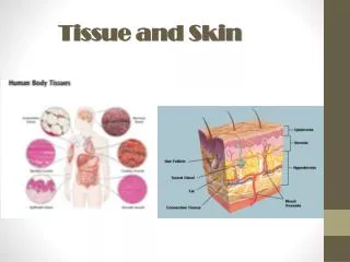
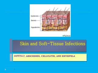
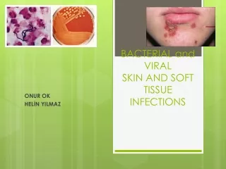
Introduction & Summary ➢ Skin and soft-tissue infections (SSTIs) variety of pathological conditions that involve the skin and underlying subcutaneous tissue, fascia, or muscle, ranging from simple superficial infections to severe necrotizing infection. 21
Introduction & Summary ➢ SSTIs manifest with : • painful • warm • Fever • erythematous skin lesions and • may also lead to purulent fluid collections • And/or necrosis of the affected tissue. 22
Introduction & Summary ➢ Risk factors for developing SSTIs (or more severe forms of SSTIs) including : oLocal factors • Chronic lymphedema • Local skin defects (e.g., tinea pedis) • Circulatory disorders: arteriovenous insufficiency, chronic edema, stasis • Peripheral neuropathy, paresis. oSystemic factors • Diabetes mellitus • Immunodeficiency (e.g., HIV infection, chemotherapy) • Chronic kidney disease (leading to, e.g., chronic edema, dialysis) • Obesity, poor nutritional status • Drug (Click...!!! ), alcohol use disorder (Click...!!!) • Older and younger age 23
Introduction & Summary o Increased exposure to pathogens • Nosocomial pathogens (e.g., prolonged hospitalization, surgery) • Water and soil exposure (e.g., sea water, Hot tub rash) (Click..!!) • Long-term intravascular devices • Trauma (e.g., open wounds, exposed fractures) ➢ Complications o Local spread of infection o Systemic involvement with fever and possible sepsis . o Spread of infection to distant sites. 24
Introduction & Summary ➢ Diagnosis is mostly clinical but some patients may require imaging or laboratory studies. ➢ Purulent infections, such as abscesses, are primarily treated with incision and drainage while nonpurulent infections (e.g., erysipelas, cellulitis) require antibiotic therapy and/or use splint for immobilization. ➢ Necrotizing soft tissue infections (NSTIs) have a high mortality rate, they are a surgical emergency and require immediate wound debridement. 25
Types of SSTI ➢ Nonbullous Nonbullousimpetigo impetigo ( (Click.... !!! Click.... !!!) ) • Multiple red papules Honey-colored crusts • Red bump => blister=>blisters rupture, ooze, form crust=> characteristic yellow scab formation ➢ Bullous impetigo ➢ vesicles or pustules that enlarge rapidly to form bullae ➢ Honey-colored varnish or crust ➢ Not painful 28
Types of SSTI ➢ Ecthyma (Click.... !!) • • • Ulceration form of impetigo , The lesions extend through the epidermis and deep into the dermis. They consist of Punched-out ulcer covered with yellow crust surrounded Painful • Ulcer with adherent crust Multiple ulcers with adherent crusts 29
Types of SSTI ➢Staphylococcal scalded skin syndrome (SSSS) : (Click ....!!!) Also known as Ritter disease is a Serious skin infection causes peeling skin over large parts of the body (superficial epidermal layer). It looks like the skin has been scalded or burned. o Caused : S.aureus produces epidermolysis exotoxin=> enters skin=> break down desmosomes between cell=> peeling skin o children under 5 years of age are at highest risk o Other risk factors include: • Weak immune system • Long-term (chronic) kidney disease or kidney failure 30
Types of SSTIs ➢ Necrotizing soft tissue infections (Click...!!!) • Mixed infection: Group A streptococci + anaerobic bacteria • Clostridial myonecrosis: Clostridium perfringens from a contaminated wound • life-threatening infection involving necrosis of the tissue. Superficial and/or deep tissue may be affected ( necrotizing cellulitis, necrotizing fasciitis, necrotizing myositis). ✓ Necrotizing cellulitis (Click...!!!) Necrotizing skin infections spread in the outer layers of skin cause infected skin and tissues to die (necrosis). The infected skin is red, warm to the touch, swollen, and gas bubbles may form under the skin. The person usually has intense pain, feels very ill, and has a high fever.
✓Necrotizing Fasciitis (Flesh-Eating Disease) (Click...!!!) (Click...!!!) • Potentially life-threatening infection => progressive destruction of deep soft tissue ( subcutaneous fat, muscle fascial) • Bacteria spread via subcutaneous tissue=> release exotoxins=> tissue destruction spreads along fascial planes ➢Common sites • Perineum ( Fournier’s gangrene): impaired gastrointestinal/urethral mucosal => spread to anterior abdominal wall gluteal muscles, scrotum, penis (male) or labia (female). Cervical ( head, neck): impaired oropharynx mucosa (dental/ odontogenic infection) => spreads to face , neck, mediastinum. • 32
✓ Necrotizing myositis (NM) (Click...!!!) • Also call necrotizing autoimmune myopathy (NAM) or immune-mediated necrotizing myopathy (IMNM) • Infection of skeletal muscle cause by clostridial myonecrosis (gas gangrene) 33
Nonpurulent SSTIs ➢ Erysipelas: superficial skin infection involving the upper dermis. ➢ Cellulitis: (Click...!!!) olocal infection of the deep dermis and subcutaneous tissue. oPossible additional features • Lymphangitis: red streaks radiating from the skin lesion and following the direction of the lymphatic vessels • Lymphadenitis: swollen, tender, regional lymph nodes. • Bullae • Purulent exudate : Cellulitis with purulent exudate is sometimes referred to as purulent cellulitis (inflamed follicles, accumulation of furuncle, abscesses cysts) 34
Nonpurulent SSTIs ➢ Erysipelas (Click...!!!) • Acute, non-necrotizing infection of upper dermis, superficial lymphatic, usually unilateral Well-defined demarcation Usually caused by streptococci most often streptococcus pyogenes. • • o R/F • • Very young/ old age Breaks in skin (e.g.. Abrasions, trauma, radiation, bite ) Lowered immunity (e.g. DMII, alcohol , DMIII, alcohol abuse, HIV .) Skin infection (e.g. tinea, impetigo) Edema ( lymphatic obstruction ) Obesity • • • • 35
Nonpurulent SSTIs ➢ Erysipelas with blisters Erysipelas with blisters A well-demarcated area of erythema on the left posterior forearm with one large blister and several smaller blisters can be seen. Some mild edema is also visible around the affected areas. These findings are consistent with erysipelas, a superficial skin infection. 36
Nonpurulent SSTIs ➢ Cellulites (Click...!!!) local infection of the deep dermis and subcutaneous tissue. o F/R of cellulities • Diabetic foot ulcers • Animal or human bites • Puncture wound • Pressure injury or pressure ulcer • Surgical wound • Water exposures • Travel-related skin infectons o Anatomic site of infection • Cellulitis of the extremities • Facial cellulitis • Neck cellulitis • Breast cellulitis • Cellulitis of abdominal wall • Cellulitis of the perineum or genitalia. 37
Nonpurulent SSTIs • Cellulitis Possible additional features ➢ Lymphangitis: red streaks radiating from the skin lesion and following the direction of the lymphatic vessel ➢ Lymphadenitis: swollen, tender, regional lymph nodes 38
Nonpurulent SSTIs • Cellulitis Possible additional features ➢ Bullae : large blisters on the skin that are filled with clear fluid. In pulmonology, it refers to an air-filled cavity that replaces lung tissue in emphysematous disease (e.g., COPD). 39
Purulent SSTIs ➢ Skin abscess : An accumulation of white-yellow pus that contains proteins, leukocytes (especially ne utrophils), bacteria, and cellular debris and is located in the dermis and subcutaneous tissue ➢ Furuncle: deep folliculitis beyond the dermis with abscess formation in the subcutaneous tissue 40
Purulent SSTIs ➢ Folliculitis (Click...!!!) localized inflammation of the hair follicle (or sebaceous glands) that is limited to the epidermis. In many cases of folliculitis, a single hair can be seen in the middle of the lesion ➢ Hot tub folliculitis: Pseudomonal folliculitis that appears 8–48 hours after exposure to contaminated water; usually a self-limiting condition that does not require antibiotic treatment 41
Purulent SSTIs ➢ Carbuncle: (Click...!!!) An infection of the hair follicle(s) that extends into the surrounding skin and deep underlying subcutaneous tissue. They typically present as an erythematous, tender, inflamed, fluctuant nodule with multiple draining sinus tracts or pustules on the surface. 42
Management of SSTI ➢ Impetigo (Click...!!!) o General: wound cleansing with antibacterial washes (e.g., chlorhexidine) o Mild nonbullous impetigo (single lesions or small are affected ) : topical antibiotics (mupirocin, retapamulin) o Bullous impetigo, ecthyma, or severe nonbullous impetigo (widespread dispersion, numerous lesions, and/or fever) • First-line treatment : first generation cephalosporins (e.g., cephalexin) or dicloxacillin • Alternative: amoxicillin-clavulanate, macrolides • If MRSA infection is confirmed or suspected : clindamycin, trimethoprim- sulfamethoxazole, doxycycline 43
Management of SSTI ➢ Staphylococcal scalded skin syndrome Staphylococcal scalded skin syndrome (Click...!!!) oHospital admission Hospital admission • Necessary in most cases • Isolation recommended • Severe cases (e.g., large areas of sloughed-off skin) may be treated in the burn unit or intensive care unit. • Outpatient treatment is possible in older children who are eating and drinking well and have minimal skin involvement. oIV IV antibiotic antibiotic • Penicillinase-resistant penicillins are the drug of choice: nafcillin, oxacillin • Vancomycin: in areas with high community in patients who do not respond to treatment) oSupportive care Supportive care • Fluid rehydration as indicated • Supportive skincare: emollients, covering sloughed-off skin • NSAIDs as indicated for pain and fever community- -acquired MRSA acquired MRSAprevalence (or 44
Management of SSTI ➢Necrotizing soft tissue infections Necrotizing soft tissue infections o Surgical exploration and Surgical exploration anddebridement • Extensive exploration with surgical debridement (removal of necrotic tissue) • Obtain deep tissue samples for Gram stain, cultures, and histopathology. o Other treatments may include Other treatments may include: • Skin grafts after the infection goes away to help your skin heal and look better • Amputation if the disease spreads through an arm or leg o Antibiotic therapy Antibiotic therapy after blood culture after blood culture • Broad-spectrum intravenous combination therapy with one of the following (MRSA coverage): • Daptomycin • Linezolid • Vancomycin • PLUS one of the following: • A carbapenem (e.g., meropenem) • Piperacillin/tazobactam • A fluoroquinolone (e.g., ciprofloxacin) PLUS metronidazole • Ceftriaxone PLUS metronidazole debridement 45
Differential diagnoses • Dermatological conditions Dermatological conditions • Acne vulgaris • Atopic dermatitis • Hidradenitis suppurativa • Vascular malformations • Viral infection Viral infections s • HSV infection (e.g., herpetic whitlow, eczema herpeticum) • Shingles • Bacterial infections Bacterial infections • Lymphogranuloma venereum • Cat scratch disease • Verruga peruana • Botryomycosis • Fungal infections Fungal infections (e.g., sporotrichosis, kerion) • Parasitic infections Parasitic infections (e.g., scabies, myiasis, larva migrans) 48
References ➢ Skin and soft tissue infections - AMBOSS ➢ https://www.uptodate.com/contents/skin-and-soft-tissue-infection-cellulitis-beyond-the- basics ➢ https://emedicine.medscape.com/article/1830144- overview?icd=login_success_gg_match_norm ➢ https://wjes.biomedcentral.com/articles/10.1186/s13017-018-0219-9 ➢ https://academic.oup.com/cid/article/59/2/e10/2895845 ➢ https://www.osmosis.org/notes/Skin_and_soft_tissue_inflammation_and_infections#page-5 ➢ https://my.clevelandclinic.org/health/diseases/15134-impetigo ➢ https://dermnetnz.org/topics/staphylococcal-scalded-skin-syndrome ➢ https://medlineplus.gov/ency/article/001443.htm ➢ https://www.merckmanuals.com/home/skin-disorders/bacterial-skin-infections/erysipelas 49
Thank You for your attention 50