Download
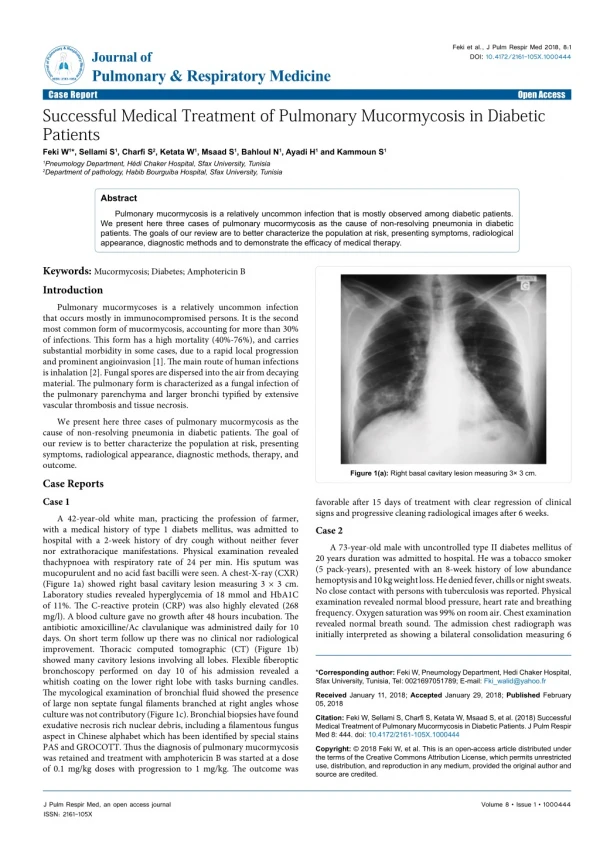
1 / 6
60 likes | 72 Views
The purpose of the conducted study was to determine which of the different types of statins ensure better control of biological markers in diabetic patients and which of the lipid molecules studied, could be the first choice of lipid-lowering therapy in people suffering from diabetes. The measurement of blood glucose levels depending on the type of statin used was another target of this research. Dyslipidaemia was a major risk factor for cardiovascular complications in people with diabetes. It was found that atorvastatin was the most effective statin in controlling dyslipidemia in diabetic, because has ensured optimum control of HDL, LDL -cholesterol, triglycerides, and glycemic values, an effect which was resulted from the particular chemical structure of atorvastatin. Atorvastatin was discovered to be the best predictor in diabetes mellitus type 2 treatment, with a sensitivity of about 78% and specificity of 45%., the highest values compared to Rosuvastatin and Simvastatin. Area Under the Curve (AUC = 0.610; AUC >0.600).

E N D
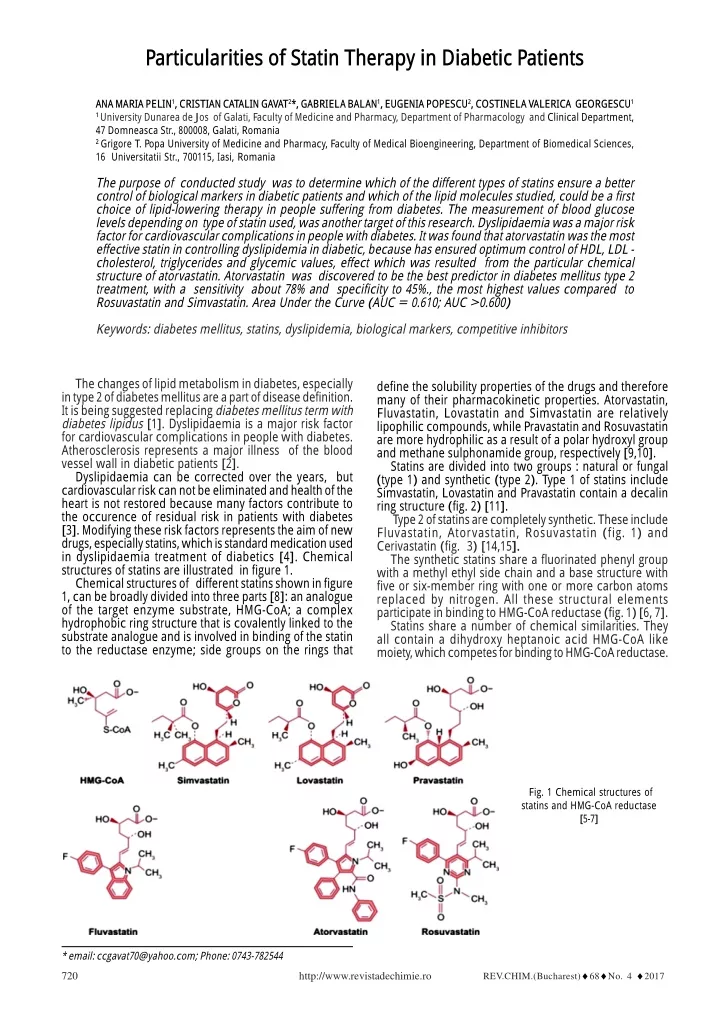
Particularities of Statin Therapy in Diabetic Patients ANA MARIA PELIN1, CRISTIAN CATALIN GAVAT2*, GABRIELA BALAN1, EUGENIA POPESCU2, COSTINELA VALERICA GEORGESCU1 1 University Dunarea de Jos of Galati, Faculty of Medicine and Pharmacy, Department of Pharmacology and Clinical Department, 47 Domneasca Str., 800008, Galati, Romania 2 Grigore T. Popa University of Medicine and Pharmacy, Faculty of Medical Bioengineering, Department of Biomedical Sciences, 16 Universitatii Str., 700115, Iasi, Romania The purpose of conducted study was to determine which of the different types of statins ensure a better control of biological markers in diabetic patients and which of the lipid molecules studied, could be a first choice of lipid-lowering therapy in people suffering from diabetes. The measurement of blood glucose levels depending on type of statin used, was another target of this research. Dyslipidaemia was a major risk factor for cardiovascular complications in people with diabetes. It was found that atorvastatin was the most effective statin in controlling dyslipidemia in diabetic, because has ensured optimum control of HDL, LDL - cholesterol, triglycerides and glycemic values, effect which was resulted from the particular chemical structure of atorvastatin. Atorvastatin was discovered to be the best predictor in diabetes mellitus type 2 treatment, with a sensitivity about 78% and specificity to 45%., the most highest values compared to Rosuvastatin and Simvastatin. Area Under the Curve (AUC = 0.610; AUC >0.600) Keywords: diabetes mellitus, statins, dyslipidemia, biological markers, competitive inhibitors The changes of lipid metabolism in diabetes, especially in type 2 of diabetes mellitus are a part of disease definition. It is being suggested replacing diabetes mellitus term with diabetes lipidus [1]. Dyslipidaemia is a major risk factor for cardiovascular complications in people with diabetes. Atherosclerosis represents a major illness of the blood vessel wall in diabetic patients [2]. Dyslipidaemia can be corrected over the years, but cardiovascular risk can not be eliminated and health of the heart is not restored because many factors contribute to the occurence of residual risk in patients with diabetes [3]. Modifying these risk factors represents the aim of new drugs, especially statins, which is standard medication used in dyslipidaemia treatment of diabetics [4]. Chemical structures of statins are illustrated in figure 1. Chemical structures of different statins shown in figure 1, can be broadly divided into three parts [8]: an analogue of the target enzyme substrate, HMG-CoA; a complex hydrophobic ring structure that is covalently linked to the substrate analogue and is involved in binding of the statin to the reductase enzyme; side groups on the rings that define the solubility properties of the drugs and therefore many of their pharmacokinetic properties. Atorvastatin, Fluvastatin, Lovastatin and Simvastatin are relatively lipophilic compounds, while Pravastatin and Rosuvastatin are more hydrophilic as a result of a polar hydroxyl group and methane sulphonamide group, respectively [9,10]. Statins are divided into two groups : natural or fungal (type 1) and synthetic (type 2). Type 1 of statins include Simvastatin, Lovastatin and Pravastatin contain a decalin ring structure (fig. 2) [11]. Type 2 of statins are completely synthetic. These include Fluvastatin, Atorvastatin, Rosuvastatin (fig. 1) and Cerivastatin (fig. 3) [14,15]. The synthetic statins share a fluorinated phenyl group with a methyl ethyl side chain and a base structure with five or six-member ring with one or more carbon atoms replaced by nitrogen. All these structural elements participate in binding to HMG-CoA reductase (fig. 1) [6, 7]. Statins share a number of chemical similarities. They all contain a dihydroxy heptanoic acid HMG-CoA like moiety, which competes for binding to HMG-CoA reductase. Fig. 1 Chemical structures of statins and HMG-CoA reductase [5-7] * email: ccgavat70@yahoo.com; Phone: 0743-782544 REV.CHIM.(Bucharest)♦68♦No. 4 ♦2017 720 http://www.revistadechimie.ro
Fig. 5. Fluorophenyl group of type 2 statins – Fluvastatin [20] Fig. 2. Chemical structures of natural statins (type 1) [12, 13] 3-hydroxy-3-methylglutaryl-CoA (HMG-CoA) reductase inhibitors (statins) act by blocking the HMG-CoA reductase enzyme, which catalyzes the rate-limiting step in de novo cholesterol synthesis. All statins are competitive inhibitors of HMG-CoA reductase with respect to the binding substrate, HMG-CoA, but not for that of the coenzyme NADPH, suggesting that their HMG-CoA-like moieties bind to the HMG-CoA-binding portion of the enzyme active site [18]. Comparison of the statin-enzyme complexes revealed subtle differences in their modes of binding. An additional hydrogen bond was found in Atorvastatin and Rosuvastatin enzyme complexes, along with a polar interaction unique to Rosuvastatin, such that Rosuvastatin has the most binding interactions with HMG-CoA reductase of all the statins [18,20]. The ring system consisted of a hydrophobic complex structure is covalently linked to the pharmacophore, which is involved in interactions with HMG-CoA reductase [19]. It has been shown that HMG-CoA reductase is stereoselective and as a result, all the statins should have chiral 1carbon atoms C3 and C5 in their pharmacophore. Statins pharmacophore inhibits HMG-CoA reductase [21]. Statins are different each from another by the hydrophobic ring and their substituents, covalently linked to HMG-like entity. Structural differences of statins affect their pharmacological properties [22]. These differences of structure are represented by the affinity to HMG-CoA site activity, the rate of liver input cell, compared to extrahepatic cells, bioavailability, metabolism, excretion, half- life time of statins [23]. Statins group has a hydrophobic state, but Rosuvastatin becomes hydrophilic due to the fact that a sulfonamide ring is present in its structure, provided with a low lipophilicity. Sulfonamide ring gives a more intense interaction with HMG-CoA - reductase. As a result, rosuvastatin has a higher afinity towards HMG-CoA reductase compared to other statins and so it has a greater effect on LDL-cholesterol lowering [10]. Statins lipophilicity is considered important because liver selectivity is in direct relationship with lipophilicity. As higher statins lipophilicity is, that much extra-hepatic tissues penetration more powerfull are, while hydrophobic statins often penetrate liver tissue [23,24]. Atorvastatin has a unique chemical structure , long plasma half-life and liver selectivity which explains high LDL-lowering potency than other inhibitors of HMG-CoA. It is a heptanoic pyrrole derivative and a synthetic LDL- lowering cholesterol. This drug increase liver receptors number, by modulating the immune response in major histocompatibility complex supression [25]. Bulky hydrophobic statin substituents might have a difficult binding relationship with HMG-CoA but conformational HMG-CoA-reductase flexibility leads to create a hydrophobic binding pocket near the active site. HMG-like functional group on statins is twisted around O5 hydroxyl group, which allows binding with HMG-CoA through a narrow area [14, 26]. Fig. 3 Chemical structure of Cerivastatin [16] The fungal statins all have a naphthalene-based ester structure (fig. 1 and fig. 2) Statin drugs inhibit HMG-CoA reductase in a competitive, dose-dependent, and reversible manner. Their structure is based on a hydrophobic ring system that is responsible for the binding to the HMG-CoA reductase in a manner that prevents binding of the natural substrate [17-19]. There is a relative variety of rings in the structure of the type 2 statins as it may be an indol ring (Fluvastatin), a pyrrol ring (Atorvastatin) or a pyrimidine ring (Rosuvastatin) [19] (fig. 1). At the moment, Rosuvastatin is considered to be the most efficient statin as it, due to its structure, enters into additional hydrogen binding interactions that increase the binding to the HMG-CoA reductase [18,19]. One of the main differences between type 1 and type 2 of statins is to replace statin type 2-fluorophenyl functional group (fig. 5) with butyryl radical found in statin 1 type group (fig. 4) [13-15]. These specific groups are responsible for additional polar interactions which creates a tighter binding with HMG reductase enzyme. Functionally, ethyl methyl group attached to the center ring of type 2 statins replaces decalin group present in type 1 statins. Butyryl group of statins type 1 occupies a similar region as fluorophenyl group present in Type 2 [15]. Fig. 4 Butyryl group of type 1 statins – Lovastatin [20] Statins are HMG-CoA reductase inhibitors, provided with inhibition constant values in the nanomolar range that effectively lower serum cholesterol levels and are widely prescribed in the treatment of hypercholesterolemia. Statins occupy a portion of the binding site of HMG-CoA, thus blocking access of this substrate to the active site. Near the carboxyl terminus of HMG-CoA reductase (HMGR), several catalytically relevant residues are disordered in the enzyme-statin complexes.If these residues were not flexible, they would sterically hinder statin binding [20]. REV.CHIM.(Bucharest)♦68♦No. 4 ♦2017 http://www.revistadechimie.ro 721
Food intake has a variable effect on statins absorption; Lovastatin is more effectively absorbed when taken along with food, whereas the bioavailability of Atorvastatin, Fluvastatin and Pravastatin is decreased. No such effect is apparent for Simvastatin or Rosuvastatin [18, 27-30]. Statins are predominantly metabolized by the cytochrome P450 (CYP450) family of enzymes, composed of over 30 isoenzymes. Statins exists in two forms: lactone (inactive form) and hydroxy acid open ring (assets) Simvastatin and Lovastatin in lactone form are prodrugs, further processed in active metabolites. On other hand, statins in β hydroxy acid form exits already in their active state and hold two hydroxyl grups in a alkyl chain β and δ positions with respect of carboxylic acid group [14, 31- 33]. One of the most important statins, Atorvastatin, must be administered in diabetic patients which were provided with a history of cardiovascular events. This treatment could be a good option but no more than 40 mg/ day administered doses of Atorvastatin[34]. Statin treatment was recommended to diabetic patients to be compulsory, because normalization of lipid levels could effective reduce the risk of stroke and major cardiovascular events, So, it was recommended a continous statin treatment, even after returning to normal lipid values, in diabetic patients case [35-37]. Statistical study. Some important statistic parameters were calculated: average values, linear regression parameters (r, R2 values), confidence interval (± 95%), p value to establish statistical differences between studied groups. ROC curve and allows to create a complete sensitivity/ specificity report. The ROC curve is a fundamental tool for diagnostic test evaluation. The area under the ROC curve (AUC) is a precise measure of how well a parameter can distinguish between two diagnostic groups (diseased/ normal) [39, 40]. In this research, ROC Curve was plotted and Area Under the Curve (AUC) was calculated, together with standard error and asymptotic confidence 95% interval (fig. 9), which completely described the sensitivity and specificity values of three studied statins. Results and discussions Analysis of biological markers into study onset in accordance with statins therapy From blood glucose analysis into study onset it was found that glucose level was significantly reduced in diabetic patients treated with Atorvastatin : 95.88 mg/dL (figure 6); p = 0.034. Glucose level was the most increased in patients who received Simvastatin treatment: 112.33 mg/ dL. Patients who have had Rosuvastatin treatment, presented a medium blood glucose level : 103.70 mg/dL (fig. 6) Experimental part It was studied a group of 50 type 2 diabetic patients diagnosed with dyslipidemia, under treatment with lipid- lowering therapy, which have been registered in the records of the diabetes and nutrition diseases section within Galati Hospital. They received different types of statins. Lipid- lowering treatment was evaluated and analized, in correlation with biological markers represented by cholesterol, HDK-cholesterol,LDL-cholesterol, triglycerides. All measured values were watched in close relationship with glycemia values a jeun (taken before meals) [36- 38]. According to lipid and glycemic parameters values obtained from initial assessment, statin type has changed in patients with diabetes mellitus. After 6 months period, biological parameters values such as cholesterol, HDL- cholesterol, LDL-cholesterol, triglycerides and glucose were reassessed. Obtained data were processed taking account of the limit values for these biomarkers: blood glucose, 70 – 110 mg/dL, triglycerides 150 mg/dL, cholesterol > 200 mg/dL, LDL-cholesterol 100 mg/dL. Doses of statins were provided with average values in studied patients: Simvastatin 40 mg/day, Rosuvastatin 10 mg/day, Atorvastatin 20 mg/day. Gender distribution showed a slightly higher frequency of women (52%), sex ratio F/M = 1.1: 1. Patient age ranged from 47 to 85 years old with a coefficient of variance 13.8%. Average group age was about 66.32 ± 9.14 years The average age in women was slightly higher than average age in males (64.46 years vs. 68.04 years), which indicates homogeneity of age by gender (p = 0.169). Before research starting, patients received three statins about 86% of them, as follows: Atorvastatin 32%, Rosuvastatin 16%, Simvastatin 38%. It was calculated mean blood glucose values colected from diabetic patients, triglycerides, HDL-cholesterol and LDL-cholesterol (mg/dL) assigned to Atorvastatin, Rosuvastatin and Simvastatin and three graphs which describe the dependency of calculated parameters according to statin type were plotted (figs. 6-8). Fig. 6. Mean blood glucose values at study onset according to statin therapy Fig. 7. Mean Triglycerides values at study onset according to statin therapy Fig. 8. Mean HDL-cholesterol values at study onset according to statin therapy The lowest triglycerides average value was observed in diabetic patients treated with Atorvastatin (110.97 mg/dL, p = 0.042) and the highest value was found in diabetic patients who received Rosuvastatin (183.69 mg/dL; p = 0.040) (fig. 7). Mean HDL-cholesterol values were significantly increased in diabetic patients treated with Atorvastatin and REV.CHIM.(Bucharest)♦68♦No. 4 ♦2017 722 http://www.revistadechimie.ro
Table 1 THE THERAPY INTO TWO STUDY STAGES presented the largest value (57.42 mg/dL; p = 0.024) (fig. 8), compared with Rosuvastatin and Simvastatin treatment ( 49.50 mg/dL and 49.97 mg/dL). Mean LDL-cholesterol values did not show any statistically significant differences (137.85 mg/dL, 120.20 mg/dL and 114.20 mg/dL respectively; p > 0.05). Currently, instituted statin therapy was focused on Atorvastatin (62%), in the expense of Simvastatin (8% of patients) or Rosuvastatin (8% of subjects). Oral antidiabetics treatment was not adjusted (table 1). At the present, the individual glucose values are directly correlated with baseline; in 42.1% of patients, elevated blood glucose levels remained (r = 0.421, R2 = 0.1775, p = 0.006). In close correlation with statin therapy, on the second review and assessment, average blood glucose level was higher in patients treated with Simvastatin, while the lowest average value was recorded in human subjects treated with Atorvastatin (p = 0.005). Individual values of triglycerides in the present are in direct correlation to baseline, over 69% of patients assumed higher values at the second assessment (r = 0.693, R2 = 0.4796, p = 0.001). Depending on statin therapy, the highest triglycerides value was recorded in diabetic patients treated with Atorvastatin (153.59 mg/dL) and the lowest value in those trated with Simvastatin (110.89 mg/ dL; p = 0.049). At the second assessment, singular values of HDL- cholesterol are in a reduced correlation with baseline ( r = 0.194, R2 = 0.0377, p = 0.243), but not significant in statistical terms . Individual values of LDL-cholesterol are directly correlated with baseline values; in 31% of patients higher levels of LDL-cholesterol were recorded ( r = 0.310, R2 = 0.0959, p = 0.062). In studied cases, the treatment with Atorvastatin has proven to be a good predictor of a favorable outcome. Areas Under the Curve – AUC = 0.610, (for a confidence interval of C.I = 95%). was calculated for patients treated with Atorvastatin. This value was ranged from 0.447 to 0.772. (fig. 9), with a sensitivity of 78% and a specificity to 45%. AUC was determined also for patients treated with other statins (Rosuvastatin and Simvastatin), which do not appear to be positive predictors in diabetes mellitus type 2 therapy and healing (AUC > 0.600) (fig. 9). Conducted statistical analysis revealed that Atorvastatin is the most effective statin drug which controls dyslipidaemia in diabetes mellitus type 2, because it ensured optimum control of HDL-cholesterol, LDL- cholesterol, triglycerides but yet in a lesser extent of total cholesterol. Fig. 9. ROC Curve - Atorvastatin specificity REV.CHIM.(Bucharest)♦68♦No. 4 ♦2017 http://www.revistadechimie.ro 723
Simvastatin ranked last place as treatment option in diabetic patient. In fact, there were some experiments which showed the important protection vascular role, especially at very high administered doses. 9. MCTAVISH, D., SORKIN, E.M, Pravastatin A review of its pharmacological properties and therapeutic potential in hypercholesterolaemia, Drugs, 42, nr. 1, 1991, p. 65-89; 10. MCTAGGART, F, BUCKET, L, DAVIDSON, R., Preclinical and clinical pharmacology of Rosuvastatin, a new 3-hydroxy-3-methylglutaryl coenzyme A reductase inhibitor, Am. J. Cardiol., 87, (Suppl. B), 2001, p. 28-32. 11. MICHAEL, S., Chemical, pharmacokinetic and pharmacodynamic properties of statins: an update, Fundam. Clin. Pharmacol. 19, 2005, p. 117–125. 12. JOSEPH, LEONARD, D., https://jleonarddixon.com/page/3/, Science: Unintended Consequences; Ancel Keys, Cholesterol And The Transition To An Obese Society; Part XV, Discover Of The Statin Drugs, Rollback Of LDL And Protection From CHD (Coronary Heart Disease), published in Science, Wonderful World of Lipids (AKA Fat), 31 May 2014, p.3. 13. RAMAKRISHNA, N., KOTESHWARA, M., VISHWOTTAM, K., Chromatography–mass spectrometry methods for the quantitation of stains in biological samples, J ournal of Pharmaceutical and Biomedical Analysis 44. nr. 2, 2007, 379-87. 14. BILJANA, N., ANA, M., MIRANDA, S., In: Chromatography - The Most Versatile Method of Chemical Analysis, 17: A Review of Current Trends and Advances in Analytical Methods for Determination of Statins: Chromatography and Capillary Electrophoresis, INTECH, World’s largest Science, Technology & Medicine Open Access book publisher, edited by Leonardo de Azevedo Calderon, 2012, p. 385-418. 15. ISTVAN, E.S., DEISENHOFER, J., Structural mechanism for statin inhibition of HMG-CoA reductase, Science 292, 2001, p. 1160–1164. 16.***International Nonproprietary Names for Pharmaceutical Substances (INN). Recommended International Nonproprietary Names (Rec. INN): List 36 (PDF). World Health Organization. 1996. p. 142. Retrieved 29 November 2016. 17. DALAL, DAHAM, AL-O., and LADISLAV, N. , Dyslipidemias and Role Of Statins In Their Therapy, Research Journal of Pharmaceutical, Biological and Chemical Sciences, 6, nr. 1, 2015, p. 449-460; 18. SRINIVASA, RAO, K., PRASAD, T., MOHANTA, G., P ., MANNA, P .,K., An Overview of Statins as Hypolipidemic Drugs, Int . J. Pharm. Sci. Drug. Res., 3, 2011, p. 178-183; 19. GAZZERRO, P ., PROTO, M., GANGEMI, G., MALFITANO, A., M., CIAGLIA, E., PISANTI, S. et al. , Pharmacological actions of statins: a critical appraisal in the management of cancer, Pharmacol. Rev., 64, 2012, p. 102-146. 20.EVA, S.I., JOHANN, D., Structural Mechanism for Statin Inhibition of HMG-CoA Reductase, Science , 292, nr. 5519, 2001, p. 1160-1164. 21. ROCHE, V., F., Teachers’ topics: antihyperlipidemic statins: a self- contained, clinically relevant medicinal chemistry lesson, Am. J. Pharm. Educ. 69 , 2005, p. 546–560. 22. FERGUS, McT., Comparative pharmacology of rosuvastatin, Atheroscler. Suppl., 4, 2003, p. 9–14. 23. LAURENCE, L.,B., Bruce, A.,C., Björn, C.,K.,, In: Goodman & Gilman’s: The Pharmacological Basis of Therapeutics, 12 Edition,, 31: Drug Therapy for Hypercholesterolemia and Dyslipidemia, Thomas P . Bersot Editor,, McGraw-Hill Ed., New York, 2011, p. 876-912. 24. WHITE, C.,M., A review of the pharmacologic and pharmacokinetic aspects of rosuvastatin, J. Clin. Pharmacol. , 42 , 2002, p. 963–970. 25. PFEFFERKORN, J., A., SONG, Y., SUN, K.,L., MILLER, S., R., TRIVEDI, B., K., CHOI, C., SORENSON, R.,J., BRATTON, L.,D., UNANGST, P .C., LARSEN, S.,D. et al., Design and synthesis of hepatoselective, pyrrole- based HMG-CoA reductase inhibitors, Bioorg. Med. Chem Lett., 17, 2007, p. 4538-4544. 26. MICHAEL, S., Chemical, pharmacokinetic and pharmacodynamic properties of statins: an update, Fundam. Clin. Pharmacol., 19, nr. 1, 2005, p. 117-125. 27. GARNETT, W.,R., Interactions with hydroxymethylglutaryl- coenzyme A reductase inhibitors. Am. J. Health Syst. Pharm. 52, nr.15,1995, p.1639- 1645. Conclusions Dyslipidaemia was a major risk factor for cardiovascular complications in people with diabetes mellitus type 2. Atorvastatin has been proven to be the most effective statin drug which ensured best control of dyslipidemia in diabetics, because it has established optimum control of HDL-cholesterol, LDL-cholesterol, triglycerides and blood glucose levels in association with hypoglycemic treatment, effect which results from Atorvastatin chemical structure. In the diabetic patients treated with Atorvastatin, blood glucose level was the lowest of the three statins : 95.88 mg/dL; p = 0.034 and presented highest statistically significant value (p = 0.05), The lowest triglycerides value was measured in diabetic patients treated with Atorvastatin (110.97 mg/dL, p = 0.042), very high statistically significant. Mean HDL-cholesterol values were significantly increased in diabetic patients treated with Atorvastatin and presented the largest value (57.42 mg/dL; p = 0.024) highest statistically significant value (p > 0.05), compared to patients treated with Rosuvastatin and Simvastatin. LDL-cholesterol values did not show any statistically significant differences in the case of three statins (137.85 mg/dL, 120.20 mg/dL and 114.20 mg/dL respectively; p > 0.05). Atorvastatin was discovered to be the best predictor in diabetes mellitus type 2 patients treatment, provided with a sensitivity about 78% and specificity to 45%., which were the most highest values compared to those of patients treated with Rosuvastatin and Simvastatin. Area Under the Curve assigned to patients treated with Atorvastatin (AUC = 0.610; AUC > 0.600). Other two statins (Rosuvastatin and Simvastatin) do not appear to be positive predictors in diabetes mellitus type 2 therapy and healing (AUC assigned to Rosuvastatin = 0.529 and AUC for Simvastatin = 0.439; AUC> 0.600). References 1. SHAFRIR, E, RAZ, I., Diabetes: mellitus or lipidus? Diabetologia, 46, nr.3, 2003, p. 433-40. 2. WANG, S.Y., HSIEH, M.C,. TU, S.T., CHUANG, C.S., New frontiers in the treatment of diabetic dyslipidemia. Rev. Diabet. Stud., 10, nr. (2- 3), 2013, p. 204-12. 3. FRUCHART, J.C, SACKS, F, HERMANS, M.P ., ASSMANN, G., BROWN, W.V., CESKA, R, CHAPMAN, M.J., DODSON, P .M., FIORETTO, P ., GINSBERG, H.,N., et al., The Residual Risk Reduction Initiative: a call to action to reduce residual vascular risk in patients with dyslipidemia, Am. J. Cardiol. 102, (Suppl.10), 2008, p. 1K–34K. 4. MARJA-RIITTA, T., Quantitative and qualitative lipoprotein abnormalities in diabetes mellitus. Diabetes. 41, (Suppl 2), 1992, p. 12–17. 5. CAMELIA, S., ANCA, S., Statins: mechanism of action and effects, J. Cell. Mol. Med., 5, nr.4, 2001, p. 378–387. 6. MCKENNEY, J.M., GANZ, P , WIGGINS, B.S., et al. In: BALLANTYNE, C.M., Editor, Clinical Lipidology A Companion to Braunwald’s Heart Disease, 2-nd Edition, PA: Saunders Elsevier, Philadelphia, 2015, p. 1- 568. 7. ALAGONA, P ., J.R., Pitavastatin: evidence for its place in treatment of hypercholesterolemia,Core Evidence, 5, 2010, p. 91-105. 8. GAW, A., PACKARD, C.J., Comparative chemistry, pharmacology and mechanism of action of the statins, In: GAW, A., PACKARD, CJ., SHEPHERD, J. (Editors), Statins. The HMG CoA reductase inhibitors in perspective, Martin Dunitz, London, 2000, pp. 49-61. REV.CHIM.(Bucharest)♦68♦No. 4 ♦2017 724 http://www.revistadechimie.ro
28. RADULOVIC, L.,L, CILLA, D.,D., POSVAR, E.,L, SEDMAN, A.,J, WHITFIELD, L.,R., Effect of food on the bioavailability of Atorvastatin, an HMG-CoA reductase inhibitor. J. Clin. Pharmacol. 35, nr. 10, 1995, p. 990-994. 29. SMITH, H.,T., JOKUBAITIS, L.,A., TROENDLE, A.,J., HWANG, D.,S., ROBINSON, W.,T., Pharmacokinetics of Fluvastatin and specific drug interactions. Am. J. Hypertens., 6, (11 pt 2), 1993, p. 375 S -382 S. 30. PAN, H.,Y., DEVAULT, A.,R., BRESCIA, D., Effect of food on Pravastatin pharmacokinetics and pharmacodynamics, Int. J. Clin. Pharmacol. Ther. Toxicol., 31, nr. 6, 1993, p. 291-294. 31. CORSINI, A., BELLOSTA, S., BAETTA, R., FUMAGALLI, R., PAOLETTI, R., BERNINI, F., New insights into the pharmacodynamic and pharmacokinetic properties of statins. Pharmacol. Ther., 84, nr.3, 1999, p. 413-428. 32. DAVIDSON, M.,H., Rosuvastatin: a highly efficacious statin for the treatment of dyslipidemia. Expert Opin. Investig. Drugs, 11, nr. 1, 2002, 125-141. 33. BOTTORFF, M., HANSTEN, P ., Long-term safety of hepatic hydroxymethyl glutaryl coenzyme A reductase inhibitors: the role of metabolism – monograph for physicians. Arch. Intern. Med., 160, nr. 15, 2000, p. 2273- 2280. 34.ROXANA, S., MOHAMMAD, A.,P ., MARJAN, A., MARYAM, T. and FARIBA, B., The effects of different doses of atorvastatin on serum lipid profile, glycemic control, and liver enzymes in patients with ischemic cerebrovascular accident, ARYA Atheroscler., 10, nr. 6, 2014, p. 298-304. 35. BISHNU, H.,S., RAJESH, T.,M., MICHAEL, G., S., MICHAEL, M.,C., SETH, S., M., DOMINIQUE,, A., M., ROGER, S., B. and MICHAEL, J., B., Diabetes Spectrum , 26, nr. 3, 2013 p. 156-164. 36. YOO-RI, CHUNG, SUNG, WOOK PARK, SHIN-YOUNG, CHOI, SEUNG, WOO KIM, KA, YOUNG MOON, JEONG, HUN KIM and KIHWANG LEE, Association of statin use and hypertriglyceridemia with diabetic macular edema in patients with type 2 diabetes and diabetic retinopathy, Cardiovasc. Diabetol. , 16, nr. 4, 2017, p.1-7. 37. DANIEL, J., M.ARSHA, Lipid Management in Patients with Type 2 Diabetes, American Health & Drug Benefits, 4, nr. 5, 2011, p. 312-322. 38. ICHIRO, KISHIMOTO, HISASHI, MAKINO, YOKO, OHATA, TAMIKO, TAMANAHA, MAYU, TOCHIYA, TOSHIHISA, ANZAI, KENGO, KUSANO, TERUO, NOGUCHI, SATOSHI, YASUDA, HISAO, OGAWA, BMJ Open Diabetes Research and Care, 3:e000137, 2015, p. 1-8. 39. CHRISTOPHER, M., F., Sensitivity, Specificity, Receiver-Operating Characteristic (ROC) Curves and Likelihood Ratios: Communicating the Performance of Diagnostic Tests, Clin. Biochem. Rev., 29, (Suppl 1), 2008, p. S83–S87. 40. KARIMOLLAH, H., T., Receiver Operating Characteristic (ROC) Curve Analysis for Medical Diagnostic Test Evaluation, Caspian J. Intern. Med., 4, nr. 2, 2013, p. 627–635. Manuscript received: 6.09.2016 REV.CHIM.(Bucharest)♦68♦No. 4 ♦2017 http://www.revistadechimie.ro 725