Download
1 / 4
40 likes | 54 Views
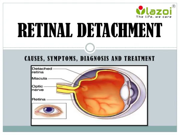
Vitreous Detachment<br>The eye is a very complex functional and anatomic organ. The retina is a thin, delicate and transparent sheet of tissue that lines the inside of the back of the eye. Directly in front of the retina is also a cavity that contains a gel called vitreous. The structure responsible for the bulk and shape of our eye is Vitreous part. It is a jelly-like body that fills the posterior chamber of the eye, giving the eyeball its round shape and keeping the retina in place against the back of the eye.

E N D
PosteriorVitreousDetachment TreatmentIn Ghatkopar,Mumbai VitreousDetachment The eye isaverycomplex functionalandanatomicorgan. Theretinaisa thin, delicateand transparentsheetoftissuethatlinestheinsideofthebackoftheeye.Directlyin frontof theretinaisalsoacavitythatcontainsagelcalledvitreous.Thestructureresponsiblefor thebulkandshapeofoureyeisVitreouspart.Itisajelly-like bodythat fillsthe posterior chamberofthe eye,givingthe eyeballitsroundshapeandkeeping theretinainplace againstthebackoftheeye. • Itismadeupofmillions oftinycollagen fibrilsalongwith groundsubstance mucopolysaccharidessuch ashyaluronicacid,whichformagel.Thevitreousismostly water,which makesup98%ofit.The collagenstrandsconnect to thesuperficiallayersof the retinaespeciallyaroundthemacula,theretinalvesselsorsitesattheretinalperiphery. • Posteriorvitreous detachment(PVD),alsoknownashyaloiddetachment,occurswhen the retinallayerandvitreousbody/posterior hyaloidmembranedissociate,with an interveningfluidcollectionformingin thesubhyaloidspace.Itisthought tobea common consequenceofaging, occurringinmorethan70%ofthepopulationoverthe ageof60 • Whoisatriskofposteriorvitreous detachment? • Theriskfactorsforvitreousdetachmentinclude: • Olderage. • Nearsightedness. • Pasteyetrauma. • PriorCataractSurgery. • Vitreousdetachmentinoneeye.
WhataresymptomsofVitreous Detachment? • Posteriorvitreous detachment (PVD)doesn’tcausepainor permanent visionloss,butyou mightexperience other symptoms.Theyinclude: • Flashes.Thesesmallflashes oflight are comparableto“seeingstars”afterhittingyour • head. Theycanlast afewseconds orminutesand tend tostop,oroccurlessoften, once detachmentiscomplete. • Floaters.Thesefloatingspotsinyourfieldofvisioncanresembletinyspecks,dust, dots,or • cobweb-likeshadows.Theytypicallyoccurin thefirstfewweeksofPosteriorvitreous detachment(PVD)and aremost noticeablewhen lookingatalightsurface,suchasawhite wallor thesky. • Cobwebeffect.Youmaybegintosee theouteredgeofthe vitreousasitseparates from • theretina.Itcanfeel like you’relookingthroughacobweb.Thisistemporary andgoesaway oncedetachment iscomplete. • HowVitreousDetachmentDevelops? • Innormaleyes,thevitreousisattachedtothesurfaceoftheretinathroughmillionsoftiny, intertwinedfibers.Yourvitreous gelismostly madeofwater. Asweage,the vitreousslowly shrinks,andthese fiberspullontheretina'ssurface.Ifthe fibersbreak,thevitreouscan shrinkfurtherandseparatefrom theretina, causinga vitreousdetachment.Most people • get Posterior vitreous detachment (PVD) at age 60 or older and it's very common after 80. Ithappenstomen andwomen equally. • Whatotherproblemscanvitreous • detachment cause? • Vitreousdetachmentcansometimesleadtomoreseriouseyeconditions: • Retinaltear.Sometimes, thevitreousfibersteara holein theretinawhentheypullaway.If you don’tgettreatmentquickly,thiscanleadtoretinaldetachment. • Retinal detachment.Sometimesvitreous detachmentpulls theentireretina awayfromits • normalpositionatthe back ofthe eye.Thiscanbea medicalemergency.Learnmoreabout retinaldetachment. • Macularhole.Sometimes vitreousdetachmenttearsa holein themacula(thepartofthe • retinathat controlsyourcentralvision).Thiscan happenbeforeorafterthevitreous detaches enough tocausefloatersorflashes oflight. Learnmore aboutmacularhole. • Macularpucker.Sometimesvitreousdetachment causesathinlayerofscartissue togrow • overthe macula. Thisusuallyhappensslowlyin themonthsoryearsaftervitreous detachment. Learnmore about macularpucker.
Theseconditionscancausevisionlossbuttreatmentmayhelppreserveyourvision.Tell youreyedoctorright awayifyou noticesymptoms ofvitreousdetachmentsotheycancheck for thesemoreseriousproblems. • Diagnosis • ARoutine eye examinationcanhelp yourdoctortospot problemslikePosteriorvitreous detachment(PVD)earlyandthatcanhelpprotectyour vision. • Yourdoctormay use specialeye dropstomakeyourpupils(theholesinthe centerofyour eyes)biggeranduseaslit-lamptest tolookfor signsofPosteriorvitreousdetachment (PVD).Thisisdonewith a microscopethatlooksthroughthe frontofyoureye. Itishelpfulin detecting, ifPosteriorvitreousdetachment(PVD)has causedbleeding, atornretina, or somethingelsethatcouldharmyoureyesight. • Your doctoralsomayuseotherteststomakesurethe gelhasn'tpulledawayfrom yourretina.Theseinclude: • Opticalcoherencetomography(OCT)- a3-D scan oftheinsideofyoureye • Ocularultrasound- atestthat usessoundwavestoshowthe insideofyoureye • Treatment • PosteriorVitreousDetachmentusuallydoesn’trequiretreatment. • Completedetachmenttypicallytakesnolongerthan threemonths.Ifyoucontinueto see floatersafterdetachmentiscomplete, discusstreatment optionswithyourdoctor. • When complications occurandyourophthalmologistrecommendstreatment,therearea number ofoptionsavailable,including: • Laserorcryosurgery:Thiscan bedoneinthe officewithno anesthesia.Yourdoctor repairsthe holesor tearsinyourretina,which preventsfurtherprogressionofthecondition. • Scleral buckle:Thisinvolvesplacing aband around theoutsideoftheeyeto • counterbalancetheforceofthevitreouspulling on theretina. Fluid isthendrained from behind yourretina,allowingit toreturn toitsproperposition. Thisisusuallydone as outpatientsurgery. • Pneumaticretinopexy:Thissurgeryissometimesdone intheoffice.Yourdoctorinjectsa • gasbubbleintothevitreousbehindyoureye.Thebubble pushesthe tearagainst the back walloftheeye andclosesit.Thenyourdoctoruseslaserorcryosurgeryto securetheretina whereitbelongs.Aftersurgery,youmayneed to keepyourheadina certain positionfora few days.Thegasbubble dissipatesovertime. • FollowupofVitreousDetachment
Once ithasbeenconfirmedthat thevitreous detachmentisisolated,follow-up examinations arerecommended atregularintervalsthereafter.The periodbetween examinations depends,ofcourse, onthepresenceofbloodin thevitreousorothersigns whichcouldincreasethelikelihoodofretinal detachment.Thusthefirstre-visitmaybe after aweekoramonth,according tothe nature ofthedetachment. ImportantReminder:Thisinformationisonlyintended to provideguidance,not a definitive medicaladvice. Pleaseconsult eyedoctoraboutyourspecificcondition.Onlya trained, experiencedboardcertifiedeyedoctorcandeterminean accuratediagnosisand proper treatment. To schedule an appointment with our experts for Posterior vitreous detachment (PVD) Treatment In Ghatkopar, please call us at +91 8451045935, +91-8451045934 or visit our clinicatAddress. Tag : best eye specialist in ghatkopar, eye clinic in ghatkopar east, Retina Detachment Treatment InGhatkopar Formoreinformation: https://www.mumbaieyecare.com/