Download
1 / 30
380 likes | 1.78k Views
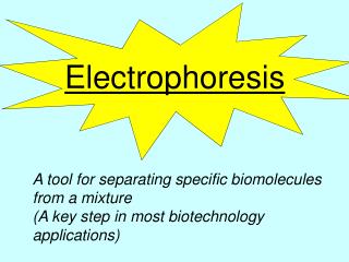
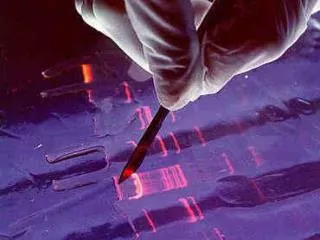
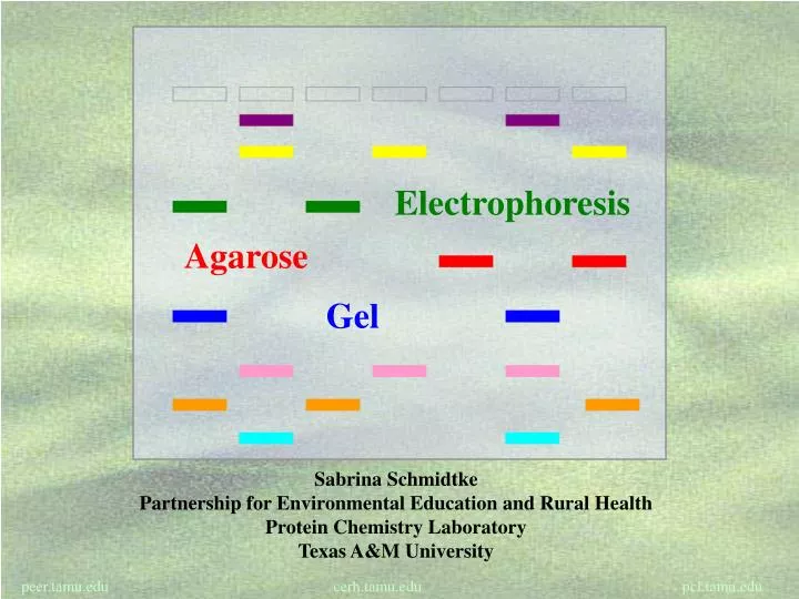
Electrophoresis. Agarose. Gel. Sabrina Schmidtke Partnership for Environmental Education and Rural Health Protein Chemistry Laboratory Texas A&M University. What is Electrophoresis?. Electrophoresis is a laboratory technique for separating mixtures of charged molecules .

E N D
Electrophoresis Agarose Gel Sabrina Schmidtke Partnership for Environmental Education and Rural Health Protein Chemistry Laboratory Texas A&M University peer.tamu.edu cerh.tamu.edu pcl.tamu.edu
What is Electrophoresis? Electrophoresis is a laboratory technique for separating mixtures of charged molecules. • Mixture:a material composed of two or more elements or parts. • Charged Molecules: a molecule (such as a protein or DNA) that has too many or too few electrons.
Positive Molecules Analyze Identify Purify Size Separation Charge Separation Mixture of Charged Molecules Negative Molecules Separation of a Mixture of Charged Molecules Charged molecules are separated based on their electrical charge and size.
Real Life Examples of Uses for Electrophoresis • Law Enforcement Agencies • Hospitals • Genetics Research
Components of Electrophoresis • Electrical Current – the flow of electric charge • Positive Electrode– the wire that collects electrons • Negative Electrode – the wire that emits electrons • Porous – containing pores, permeable to fluids and small particles • Sieve –a mesh device to filter small particles out of a mixture of larger particles.
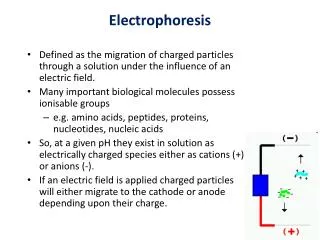
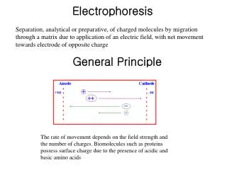
+ + - + O - - + - + N - - - - - + + - + - + + - - + - + N Negatively Charged Protein Positively Charged Peptide Positively Charged Amino Acid How Separation Occurs Electrical Charge: Many molecules (amino acids, peptides, proteins, DNA, and RNA) have naturally occurring negative and positive charges on them. The sum of these charges determines the overall charge. When introduced to an electrical current, negatively charged molecules are attracted to the positive electrode and positively charged molecules are attracted to the negative electrode.
Porous Material Proteins Entering Porous Material Smallest Move Fastest How Separation Occurs Molecule Size: The porous material is made of microscopic particles suspended in a gel. The microscopic particles attach to one another forming tunnels that act as a sieve to separate the molecules. Small molecules can move faster than large molecules.
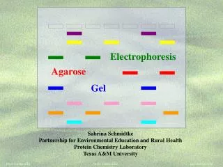
Gel Electrophoresis Gels can be made from substances such as agarose or polyacrylamide. • Agarose –a complex sugar chain from red seaweed. It is commonly used in foods (ice cream, whipped cream, and jellies) and many biological mediums. It has a large pore size good for separating large molecules quickly. • Polyacrylamide –chain of acrylic acid molecules. It is often used to make plastics and rubber. It has a small pore size good for separating small molecules slowly. *Polyacrylamide is a neurotoxin! Red Sea Weed Acrylic Acid
Overview of Gel Electrophoresis • Mix agarose or polyacrylamide powder with liquid buffer. • Pour the gel into a mold. • Place a comb in the gel to form sample wells. • Allow the gel to solidify. • Submerge the gel in a tank full of a liquid buffer. • Place the samples in the wells. • Turn on the power source. • Charged molecules will move to the oppositely charged electrode. • Turn off the power source and remove the gel. • Observe the separated molecules.
Illustration of Gel Electrophoresis - - Negative Electrode - - - - Negative Electrode - - Wells + + Positive Electrode + + + + Positive Electrode + + Before Electrophoresis After Electrophoresis
Gel Electrophoresis Experiment Edible Colors
Overview of the Experiment Purpose: To introduce the principles and terminology of electrophoresis and demonstrate the separation of food coloring dyes with agarose gel electrophoresis.
Materials List Chamber and Power Supply • Plastic dish • Slide box • Aquarium sealant • Large needle • Seizing wire • Hot glue gun and glue sticks • Nine-volt batteries • Nine-volt battery clip • Alligator clips • Scissors
Materials List Samples and Gel Preparation • Small test tubes/vials • Tube rack • Permanent marker • Transfer pipettes • Capillary tubes with bulb • Food colors • 50% glycerol solution • 2” x 3” glass slide • Well comb • Tris-Borate-EDTA Buffer (TBE) • Agarose • Graduated cylinder • Erlenmeyer flask • Balance • Microwave or hot water bath
Safety Precautions • Chemicals – Aquarium Sealant, Agarose, and TBE Buffer are all irritants. If you get them on your skin or in your eyes, rinse with water. • Electricity – do not touch the alligator clips or the buffer when the power supply is assembled and hooked up. IT WILL ELECTROCUTE YOU!!!! • Hot Objects – the hot glue gun and the agarose solution will both be hot. If you get burned, rinse the burn with cool water and seek medical attention if necessary. • Glassware – (beakers, graduated cylinders, slides) If anything is broken dispose of the glassware in an appropriate manner. • Sharps – the needle, seizing wire ends, and capillaries are all sharp. Be careful when handling these items and dispose of them properly.
Building the Electrophoresis Chamber • Place the slide box in the center of the plastic dish and trace around the edge with the permanent marker. • Extend the two long sides up one side of the plastic dish. • With the marker, place a spot near the rim of the plastic dish, about 1 cm out from each of the lines.
Building the Electrophoresis Chamber • Carefully use the large needle to punch a hole at each spot. • Cut a piece of wire about 10 cm longer than the length of the plastic dish. • Bend the wire as shown. • The wire should touch the plastic dish on both sides, but be about 1 cm off of the bottom.
Building the Electrophoresis Chamber • Put a thick bead of aquarium sealant around the rim of the slide box. • Place the slide box into the plastic dish. • Allow the aquarium sealant to dry overnight.
Building the Power Supply • Cut the battery clip in half with the scissors. Be careful not to cut the wires. • Remove one cover off each alligator clip. • Feed the battery clip wire through the cover and wrap it around the appropriate alligator clip. • Replace the cover to protect the wire connection. • Connect the batteries into a pyramid. • Connect the battery clips to the batteries
Preparing the Gel • Place agarose and TBE buffer into the Erlenmeyer flask. • Heat the flask in the microwave or a hot water bath. The flask will become very hot. • When the agarose is dissolved, there will be no more particles in the TBE Buffer. • Allow the flask to cool until you are able to touch it without burning yourself.
Preparing the Gel • Lay the glass slide on the table. • Use the binder clips to hold the comb just above the slide.
Pouring the Gel • Use a transfer pipette to place the agarose onto the slide. • Start at the comb and work your way out covering the whole slide. Pop any air bubbles immediately. • When the gel has solidified it will become opaque. • Carefully remove the comb.
Preparing and Loading the Samples • Label the test tubes with the color of food dye you will put in them. • Mix one drop of food coloring with three drops of 50% glycerol. Repeat with all samples. • Carefully place the gel in the plastic dish on top of the slide box. • Fill the chamber with TBE Buffer until the gel is covered. • Use the capillary tubes to load 10 μl of sample in each well.
Developing the Gel • Assemble the battery pyramid and connect alligator clips. • Connect the black alligator clip to the wire behind the wells and the red alligator clip to the wire in front of the wells. • DO NOT touch the buffer while the power supply is attached to the chamber! • Allow the gel to develop for at least 35 minutes. You will be able to see the dyes separate.
Observing the Gel • Disconnect the alligator clips and take apart the battery pyramid. • Carefully remove the slide with gel and lay on a paper towel. • Observe the dye separation of the individual food colors. • Compare the dye separation of the mixtures with the dye separation of the individuals All Red Blue Green Yellow
Observing the Gel • Yellow food coloring separated into yellow dye • Red food coloring separated into red dye and pink dye • Green food coloring separated into yellow dye and blue dye • Blue food coloring separated into red dye and blue dye • The mixed sample contains all of the dyes Red Blue Green Mixed Yellow
Observing the Gel • This gel was run for 120 minutes, it shows better separation of the dyes and good replication for the dyes. • The size of molecules from smallest to largest are: yellow, red, pink, and blue. Red Red Blue Blue Green Yellow Yellow Mixed
Alternative Experiments • Other Samples • Separate the food dyes used in Kool-Aid and Skittles. • Separate proteins and DNA. (will require additional materials) • pH Change • Change the pH of the buffer in the gel and the tank to observe the changes it makes on the samples. • Change the Percentage of Agarose Used • Observe how using higher/lower concentrations of agarose will change the separation of dyes.
1 2 3 4 5 Alternative Experiments Skittles 1) Grape 2) Lime 3) Lemon 4) Orange 5) Strawberry
1 2 3 4 5 6 Alternative Experiments Kool-Aid 1) Strawberry 2) Orange 3) Tropical Punch 4) Grape 5) Ice Blue Raspberry Lemonade