1 / 38
380 likes | 409 Views
Training module created for all those involved with airway management working at Liverpool University Hospitals NHS Foundation Trust, covering preventing undetected oesophageal intubation, 'shared airway' principles, basic interpretation of capnography and communication tools to challenge authority. We are aiming for this to become part of the Annual Role Specific Mandatory Training for all Anaesthetists, Intensivists, ODPs and A and E doctors in the trust.

E N D
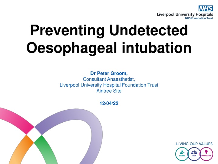
Preventing Undetected Oesophageal intubation Dr Peter Groom, Consultant Anaesthetist, Liverpool University Hospital Foundation Trust Aintree Site 12/04/22
By the end of this module you will be able to: • Understand what oesophageal intubation is and why it is important to recognise it • Understand the role capnography plays in detecting misplaced endotracheal tubes • Understand basic capnography trace patterns and when to be concerned • Be able to explain how we can work together to prevent oesophageal intubation • Be able to apply communication tools to help challenge authority where you have a concern that is not being heard
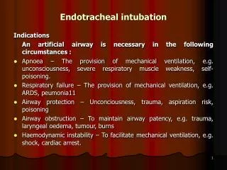
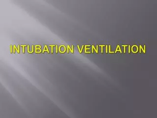
What is Oesophageal Intubation? • Oesophageal intubation is when an endotracheal tube is passed into the oesophagus instead of the trachea • The lungs will therefore NOT be ventilated • If unrecognised, worsening hypoxia results leading to cardiac arrest and ultimately death • It is RARE • It is FATAL • It is AVOIDABLE: looking at and acting on the patient’s capnography trace identifies the problem quickly • Our hospital considers it a NEVER EVENT
Why is recognising oesophageal intubation important? • NAP 4 ( the largest study of major complications of airway management ever performed) identified 9 cases of oesophageal intubation, resulting in 6 deaths. • Mrs Glenda Logsdail, a healthy 61 year old radiographer, listed for an appendicectomy, had an unrecognised oesophageal intubation. Anaesthetists misinterpreted the cause of a flat capnograph trace. She suffered a prolonged period of hypoxia before the misplaced endotracheal tube was diagnosed. She died on the 23rd August 2020 from hypoxaemic encephalopathy. • HM Coroner issued a Prevention of Future Deaths communication Mrs Glenda Logsdail
Role of Capnography • Capnography is continuous monitoring of carbon dioxide (CO2) in respiratory gases and gives a visual, breath by breath, assessment of the patient's airway and ventilation. • Capnography is AAGBI mandated standard monitoring for all patients with an artificial airway and patients receiving advanced life support • Capnography is the GOLD STANDARD to confirm correct tube placement and to detect tube misplacement. • Clinical signs e.g. chest expansion and misting of the tube are unreliable and should not be relied upon to confirm tube position instead of capnography • Capnography MUST be used for ALL tracheal intubations and continue to be used until after extubation. This is MANDATORY for every location in our hospital. • Yellow capnography waveform
CO2 TRACE = CORRECT PLACE A capnography trace from a patient with a correctly sited endotracheal tube or tracheostomy, where ventilation is occurring.
NO TRACE = WRONG PLACE • A flat capnography trace = the lungs are NOT being ventilated. • A flat trace MUST be assumed to be due to oesophageal intubation. • This applies during cardiac arrest. • The tracheal tube is in the wrong place.
Capnography patterns: The Hats and Caps tool Everyone involved in airway management must be able to recognise these four traces Hat A shows the endotracheal tube or tracheostomy is correctly placed and the patient is being ventilated normally Hat B has a sloped top indicating ventilation is occurring but with a degree of obstruction secondary to • A tracheal tube that has migrated beyond the carina into a bronchus • Bronchospasm • COPD Kerslake and Kelly. BJA Education 2017;17(5): 178-183
Capnography patterns: The Hats and Caps tool Hat C Indicatres a leak around an endotracheal tracheal or tracheostomy tube Hat D FLAT TRACE = NO VENTILATION IS OCCURING. Tube is in the wrong place Oesophageal intubation until proven otherwise It must be recognised and acted on immediately. • NO TRACE = WRONG PLACE Kerslake and Kelly. BJA Education 2017;17(5): 178-183
Capnography patterns • A: Sudden drop in CO2 trace suggests a sudden drop in lung blood flow, for example due to: • Pulmonary embolism • Drop in cardiac output • B: Flat line = oesophageal intubation. • It can take up to 6 waveforms with decreasing height to reach a flat line • This is due to some CO2 being present in the gas in the stomach particularly if there has been difficult facemask ventilation. • It is best practice to wait for 6 breath cycles after intubation before confirming the tube is in the trachea. Kerslake and Kelly. BJA Education 2017;17(5): 178-183
Other causes of a flat capnograph trace • FIRST EXCLUDE OESOPHAGEAL INTUBATION then consider other causes of a flat capnograph trace • 1. Blocked Endotracheal Tube (blood, secretions, kinked tube, patient biting) • Inability to pass a suction catheter identifies this • 2. Circuit disconnection or blockage • Visually inspect the breathing circuit. • Regaining a trace by using a self-inflating bag connected DIRECTLY to tracheal tube connectoridentifies a circuit issue quickly • 3. Water in the circuit or tubing of a side-stream capnograph • 4. Occlusion of capnograph sample line • 5. Life threatening bronchospasm
If you see a flat capnograph trace • Declare loudly there is a flat capnograph and possible oesophageal intubation • Ensure the anaesthetist is alerted • If you feel dismissed use the CUS communication tool to make sure you are heard (covered later in the module). • Increase gas flows and apply 100% Oxygen • Perform quick visual inspection of entire breathing system including valves and connections, looking for blockage or disconnection • Remember the mantra ‘IF IN DOUBT, TAKE IT OUT’. • Deflate the cuff and extubate • Face mask ventilate with 100% O2 and prepare to reintubate. • Videolaryngoscopy allows a two person visual confirmation of the tube passing through the vocal cords. • Confirm correct position of the tube using capnography for a minimum of 6 breaths. • Follow the DAS 2015 guidelines or Vortex cognitive aid If there is difficulty intubating
Capnography and cardiac arrest During cardiac arrest • If the tube is in the trachea and you are ventilating the patient, there WILL BE an attenuated capnograph trace • A flat capnograph trace means the lungs are not being ventilated and therefore the tube is in the wrong place. Assume oesophageal intubation until this is actively excluded.
SHARED AIRWAY prevents oesophageal intubation • The ‘Shared Airway’ = Anaesthetist and ODP using videolaryngoscopy and capnography together to design out oesophageal intubation • Videolaryngoscopy allows the ODP as well as anaesthetist observe the view at laryngoscopy and verify tube placement. • The shared airway is a 2 person check (anaesthetist and ODP) verbally confirming when you both see: • 1. The tube passed through the cords • 2. The circuit is connected • 3. Ventilation is commenced • 4. Capnography is confirmed for a minimum of 6 breaths • The team’s use of videolaryngoscopy and capnography together will flatten team hierarchy and improve recognition of oesophageal intubation.
Communication tools can help your voice to be heard • In a crisis, interpersonal behaviour WILL deteriorate. The person involved in a crisis situation may become task fixated and lose situational awareness. This is when other members may need to intervene if patient safety is compromised. • CUS is a tool that can be used to help challenge authority when you feel there is a patient safety issue.
Version 1.1 of this module was prepared by Dr Groom and Dr Amarasekara in Spring 2022 It is due for review April 2024 Permission was granted by Mr Richard Logsdail for the use of his wife’s photograph 12/4/22
Thankyou for completing this presentation. To complete the module you must now pass the MCQ with a score of 100% Consider reviewing the presentation before proceeding as you are only allowed 3 attempts
Q1. What is the best method for confirming endotracheal tube placement in the trachea?(select any that apply) 1. Visualizing the endotracheal tube passing through the vocal cords? 2. Visualizing “misting” or “fogging” of the endotracheal tube ? 3. Visualizing the “chest rise and fall ? 4. Confirming lung sounds with a stethoscope? 5. Confirming a waveform on the capnograph?
Q2. Immediately following intubation what is the most likely cause of an absent capnography trace?(select any that apply) 1. Anaphylaxis? 2. Cardiac arrest? 3. Low blood pressure? 4. Oesophageal intubation? 5. Bronchospasm?
Q3: Capnography during a cardiac arrest with effective chest compressions shows: 1.No wave form ? 2. A smaller amplitude, normal pattern wave form ?
Q4. Following intubation, which of these four capnograph traces shows a clear, unobstructed airway?
Q5. Which of these four capnograph traces is seen after an oesophageal intubation?
Q6. Following intubation which of these four capnograph traces is seen with bronchospasm?
Q7. Following intubation, which of these four capnograph traces is seen with a leak around the endotracheal tube?
Q8. Which of these four capnograph traces is seen when an endotracheal tube has become displaced or dislodged from the trachea ?
Q9. Which of these four capnograph traces is seen when the lungs are not being ventilated?
Q10. Concerning anaesthesia and airway management (select any that apply) • Use of capnography is mandatory standard of monitoring for intubation? • Oesophageal intubation is excluded by misting of the airway with ventilation? • Oesophageal intubation is excluded by the chest rising with ventilation? • Oesophageal intubation is excluded breath sounds heard through a stethoscope with ventilation? • Oesophageal intubation is excluded with a normal, “top hat” capnograph trace?
Q11. Which of these traces indicate oesophageal intubation? 1. 2. 3.
Q12. Which of these are causes of a flat capnograph trace. Tick all that apply • Cardiac arrest during CPR • Endotracheal tube blocked by blood • Oesophageal intubation • Patient has bitten the tube • Tracheostomy that has been inserted through a false passage • The CO2 sample line has not been connected • Malignant Hyperthermia
Q13. You are performing a 2 person check at intubation using a McGrath videolaryngoscope. Which screen shows a correctly sited endotracheal tube? A B C
Q14. You are the ODP, and you notice after intubation that there is a flat capnograph trace. What should you do first? • Attribute this to an equipment issue and secure the tube in place • Declare there is a flat capnograph to the team and alert anaesthetist • Get the Quick reference Handbook from the cupboard • Visually inspect the entire breathing system for disconnection • Connect the tube directly to a self-inflating bag
Q15 You have alerted the anaesthetist after intubation to a flat capnograph trace but they are not listening to you. What can you do next? • Use the tool CUS (I am Concerned, I am Uncertain, This is a Safety issue) • Use the tool SBAR (Situation, Background, Assessment, Recommendation) • Use the tool CUS (I am Concerned, I am Uncomfortable, This is a Safety issue) • Use the tool RSVP(Reason, Story, Vital Signs, Plan)
Q16 You use the CUS communication tool to assert yourself. Order the following statements using this tool: • ‘Dr X, I am uncomfortable, I cannot see an ETCO2 trace.’ • ‘Dr X, this needs to stop now. I am going to have to call for another anaesthetist to help.’ • ‘Dr X, I am concerned I cannot see an ETCO2 trace’ • ‘Dr X, this is now unsafe. I cannot see an ETCO2 trace and the patient saturations are dropping. I do not think this tube is in the right place. I think we need to reposition the tube.’