Download
1 / 70
700 likes | 1.15k Views
Chapter 43. The Immune System. Overview: Reconnaissance, Recognition, and Response. An animal must defend itself from the many dangerous pathogens it may encounter Two major kinds of defense have evolved: innate immunity and acquired immunity.

E N D
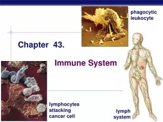
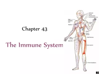
Chapter 43 The Immune System
Overview: Reconnaissance, Recognition, and Response • An animal must defend itself from the many dangerous pathogens it may encounter • Two major kinds of defense have evolved: innate immunity and acquired immunity
Innate immunity is present before any exposure to pathogens and is effective from the time of birth • It involves nonspecific responses to pathogens • Innate immunity consists of external barriers plus internal cellular and chemical defenses • Key internal defenses are macrophages and other phagocytic cells
Acquired immunity, or adaptive immunity, develops after exposure to agents such as microbes, toxins, or other foreign substances • It involves a very specific response to pathogens • Recognition is by white blood cells called lymphocytes • Some lymphocytes produce antibodies; others destroy infected cells, cancer cells, or foreign tissue
ACQUIRED IMMUNITY Slower responses to specific microbes INNATE IMMUNITY Rapid responses to a broad range of microbes External defenses Internal defenses Skin Phagocytic cells Humoral response (antibodies) Mucous membranes Antimicrobial proteins Secretions Inflammatory response Invading microbes (pathogens) Cell-mediated response (cytotoxic lymphocytes) Natural killer cells The BIG PICTURE!!!
Concept 43.1: Innate immunity provides broad defenses against infection • A pathogen that breaks through external defenses encounters innate cellular and chemical mechanisms that impede its attack
External Defenses • Skin and mucous membranes are physical barriers to entry of microorganisms and viruses • Mucous membrane cells produce mucus, a viscous fluid that traps microbes and other particles • In the trachea, ciliated epithelial cells sweep mucus and any entrapped microbes upward, preventing microbes from entering the lungs
LE 43-3 10 µm
Secretions of the skin and mucous membranes provide an environment hostile to microbes • Secretions give the skin a pH between 3 and 5, acidic enough to prevent colonization of many microbes • Skin secretions include proteins such as lysozyme, which digests bacterial cell walls
Internal Cellular and Chemical Defenses • Internal cellular defenses depend mainly on phagocytosis • White blood cells called phagocytes ingest microorganisms and initiate inflammation
Phagocytic Cells • Phagocytes attach to prey via surface receptors and engulf them, forming a vacuole that fuses with a lysosome
Macrophages, a type of phagocyte, migrate through the body and are found in organs of the lymphatic system • The lymphatic systemdefends against pathogens
Lymphatic capillary Interstitial fluid Adenoid Tonsil Blood capillary Lymph nodes Lymphatic vessel Spleen Tissue cells Peyer’s patches (small intestine) Appendix Lymphatic vessels Masses of lymphocytes and macrophages Lymph node
Antimicrobial Proteins • Proteins function in innate defense by attacking microbes directly or impeding their reproduction • About 30 proteins make up the complement system, which causes lysis of invading cells and helps trigger inflammation • Interferons provide innate defense against viruses and help activate macrophages
Pin Blood clot Pathogen Macrophage Blood clotting elements Chemical signals Phagocytic cells Phagocytosis Capillary Red blood cell Inflammatory Response • In local inflammation, histamine and other chemicals released from injured cells promote changes in blood vessels • These changes allow more fluid, phagocytes, and antimicrobial proteins to enter tissues
Natural Killer Cells • Natural killer (NK) cellsattack virus-infected body cells and cancer cells • They trigger apoptosis in the cells they attack
Invertebrate Immune Mechanisms • Many invertebrates defend against infection by many of the same mechanisms in the vertebrate innate response
Concept 43.2: In acquired immunity, lymphocytes provide specific defenses against infection • Acquired immunity is the body’s second major kind of defense • An antigen is a foreign molecule that is recognized by lymphocytes and elicits a response from them • A lymphocyte recognizes and binds to a small portion of the antigen called an epitope
Antigen- binding sites Epitopes (antigenic determinants) Antibody A Antigen Antibody B Antibody C
Antigen Recognition by Lymphocytes • Two main types of lymphocytes circulate in the blood of vertebrates: B lymphocytes (B cells) and T lymphocytes (T cells) • A single B cell or T cell has about 100,000 identical antigen receptors • All antigen receptors on a single cell recognize the same epitope SPECIFICITY IS THE WORD!!!
B Cell Receptors for Antigens • B cell receptors bind to specific, intact antigens • Secreted antibodies, or immunoglobulins, are structurally similar to B cell receptors but lack transmembrane regions that anchor receptors in the plasma membrane
Antigen- binding site Antigen- binding site Antigen- binding site Disulfide bridge V V V V Light chain Variable regions V V C C C C C C Constant regions Transmembrane region Plasma membrane b chain chain Heavy chains Disulfide bridge T cell B cell Cytoplasm of B cell Cytoplasm of T cell A B cell receptor consists of two identical heavy chains and two identical light chains linked by several disulfide bridges. A T cell receptor consists of one a chain and one b chain linked by a disulfide bridge.
T Cell Receptors for Antigens and the Role of the MHC • Each T cell receptor consists of two different polypeptide chains
T cells bind to antigen fragments that are bound to cell-surfaceproteins called MHC molecules • MHC molecules are so named because they are encoded by a family of genes called the major histocompatibility complex
Infected cells produce MHC molecules, which bind to antigen fragments and are transported to the cell surface, a process called antigen presentation • A nearby T cell can then detect the antigen fragment displayed on the cell’s surface • Depending on their source, peptide antigens are handled by different classes of MHC molecules
Class I MHC molecules are found on almost all nucleated cells of the body • They display peptide antigens to cytotoxic T cells
Dendritic cells have lots of “arms” – they act as antigen-presenting cells • Class II MHC molecules are located mainly on dendritic cells, macrophages, and B cells • They display antigens to helper T cells
Lymphocyte Development • Lymphocytes arise from stem cells in bone marrow • Newly formed lymphocytes are alike but later develop intoB cells or T cells, depending on where they mature
Bone marrow Thymus Lymphoid stem cell B cell T cell Blood, lymph, and lymphoid tissues (lymph nodes, spleen, and others) B T
Generation of Lymphocyte Diversity by Gene Rearrangement • Random, permanentgene rearrangementforms functional genesencodingthe B or T cell antigen receptor chains
V4–V39 DNA of undifferentiated B cell V1 V40 J1 J4 J5 V3 J3 V2 J2 C Intron Deletion of DNA between a V segment and J segment and joining of the segments DNA of differentiated B cell V1 V3 V2 J5 C Intron Functional gene Transcription of resulting permanently rearranged, functional gene J5 V3 C Intron pre-mRNA RNA processing (removal of intron; addition of cap and poly (A) tail) J5 V3 C mRNA Poly (A) Cap Translation B cell receptor Light-chain polypeptide V C Variable region Constant region B cell Let’s not go here…
Testing and Removal of Self-Reactive Lymphocytes • As B and T cells mature in the bone and thymus, their antigen receptors are tested forself-reactivity • Lymphocytes with receptors for antigens that are already in the body are destroyed by apoptosis or rendered nonfunctional
Clonal Selection of Lymphocytes • In a primary immune response, binding of antigen to a mature lymphocyte induces the lymphocyte’s proliferation and differentiation • This process is called clonal selection • Clonal selection of B cells generates a clone of short-lived activated effector cells and a clone of long-lived memory cells Animation: Role of B Cells
Antigen molecules B cells that differ in antigen specificity Antigen receptor Antibody molecules Clone of plasma cells Clone of memory cells
104 103 Antibodies to A Antibody concentration (arbitrary units) 102 Antibodies to B 101 100 21 7 0 28 49 14 35 56 42 Time (days) • In the secondary immune response, memory cells facilitate a faster, more efficient response
and teens too! CDC has all the info about immunizations
Concept 43.3: Humoral and cell-mediated immunity defend against different types of threats • Humoral immune response involves activation and clonal selection of B cells, resulting in production of specific secreted antibodies • Cell-mediated immune response involves activation and clonal selection of cytotoxic T cells
Cell-mediated immune response Humoral immune response First exposure to antigen Antigens displayed by infected cells Antigens engulfed and displayed by dendritic cells Intact antigens Activate Activate Activate Secreted cytokines activate B cells Cytotoxic T cell Helper T cell Gives rise to Gives rise to Gives rise to Active and memory helper T cells Memory cytotoxic T cells Active cytotoxic T cells Plasma cells Memory B cells Defend against infected cells, cancer cells, and transplanted tissues Secrete antibodies that defend against pathogens and toxins in extracellular fluid Another BIG PICTURE to put it all together
Helper T Cells: A Response to Nearly All Antigens • A surface protein called CD4 binds the class II MHC molecule • This binding keeps the helper T cell joined to the antigen-presenting cell while activation occurs • Activated helper T cells secrete cytokines that stimulate other lymphocytes Animation: Helper T Cells
Peptide antigen Cytotoxic T cell Dendritic cell Class II MHC molecule Cell-mediated immunity (attack on infected cells) Helper T cell Bacterium TCR Humoral immunity (secretion of antibodies by plasma cells) CD4 Dendritic cell B cell Cytokines
Cytotoxic T Cells: A Response to Infected Cells and Cancer Cells • Cytotoxic T cells make CD8, a surface protein that greatly enhances interaction between a target cell and a cytotoxic T cell • Binding to a class I MHC complex on an infected cell activates a cytotoxic T cell and makes it an active killer • The activated cytotoxic T cell secretes proteins that destroy the infected target cell
Released cytotoxic T cell Cytotoxic T cell Perforin Cancer cell Granzymes TCR Apoptotic target cell CD8 Class I MHC molecule Pore Target cell Peptide antigen Cytotoxic T cell Animation: Cytotoxic T Cells
Antibody Classes • The five major classes of antibodies, or immunoglobulins, differ in distribution and function • They are called IgA, IgD, IgE,IgG and IgM
B Cells: A Response to Extracellular Pathogens • Activation of B cells is aided by cytokines and antigen binding to helper T cells • Clonal selection of B cells generates antibody-secreting plasma cells, the effector cells of humoral immunity
Macrophage Bacterium Peptide antigen B cell Class II MHC molecule Secreted antibody molecules Clone of plasma cells TCR CD4 Endoplasmic reticulum of plasma cell + Cytokines Helper T cell Activated helper T cell Clone of memory B cells Why is there lots of ER in this cell? LE 43-17
IgG (monomer) IgM (pentamer) Most abundant lg class in blood; also present in tissue fluids First lg class produced after initial exposure to antigen; then its concentration in the blood declines Only lg class that crosses placenta, thus conferring passive immunity on fetus Promotes neutralization and agglutination of antigens; very effective in complement activation (see Figure 43.19) Promotes opsonization, neutralization, and agglutination of antigens; less effective in complement activation than lgM (see Figure 43.19) J chain The Immunoglobulin Family Opsonization = marking antigen for destruction/phagocytosis
IgA (dimer) Present in secretions such as tears, saliva, mucus, and breast milk IgD (monomer) Present primarily on surface of naive B cells that have not been exposed to antigens IgE (monomer) Provides localized defense of mucous membranes by agglutination and neutralization of antigens (see Figure 43.19) Triggers release from mast cells and basophils of histamine and other chemicals that cause allergic reactions (see Figure 43.20) J chain Acts as antigen receptor in antigen-stimulated proliferation and differentiation of B cells (clonal selection) Presence in breast milk confers passive immunity on nursing infant Transmembrane region Secretory component
Antibody-Mediated Disposal of Antigens • The binding of antibodies to antigens is also the basis of antigen-disposal mechanisms • Microbes are eliminated by phagocytosis and complement-mediated lysis Animation: Antibodies