Download
1 / 3
30 likes | 37 Views
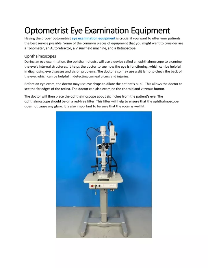
Having the proper optometrist eye examination equipment is crucial if you want to offer your patients the best service possible. Some of the common pieces of equipment that you might want to consider are a Tonometer, an Autorefractor, a Visual field machine, and a Retinoscope.<br>Ophthalmoscopes<br>During an eye examination, the ophthalmologist will use a device called an ophthalmoscope to examine the eye's internal structures. It helps the doctor to see how the eye is functioning, which can be helpful in diagnosing eye diseases and vision problems. The doctor also may use a slit lamp to check the b

E N D
Optometrist Eye Examination Equipment Optometrist Eye Examination Equipment Having the proper optometrist eye examination equipment is crucial if you want to offer your patients the best service possible. Some of the common pieces of equipment that you might want to consider are a Tonometer, an Autorefractor, a Visual field machine, and a Retinoscope. Ophthalmoscopes Ophthalmoscopes During an eye examination, the ophthalmologist will use a device called an ophthalmoscope to examine the eye's internal structures. It helps the doctor to see how the eye is functioning, which can be helpful in diagnosing eye diseases and vision problems. The doctor also may use a slit lamp to check the back of the eye, which can be helpful in detecting corneal ulcers and injuries. Before an eye exam, the doctor may use eye drops to dilate the patient's pupil. This allows the doctor to see the far edges of the retina. The doctor can also examine the choroid and vitreous humor. The doctor will then place the ophthalmoscope about six inches from the patient's eye. The ophthalmoscope should be on a red-free filter. This filter will help to ensure that the ophthalmoscope does not cause any glare. It is also important to be sure that the room is well lit.
Retino Retinoscopes scopes During an optometrist eye examination, the doctor uses a retinoscope to determine if your eyes need corrective lenses. This is an easy and painless procedure that allows the doctor to see inside the eye. The retinoscope is a handheld device that moves a beam of light across the pupil. The result is an accurate measurement of your lens power. The retinoscope may be used manually or with an automated machine. If you're in the market for a new pair of glasses, a retinoscope can be the best way to find the right prescription. In most cases, a retinoscope will be used for a routine eye exam. This is a good option for patients with special needs, and it can be performed outside of a traditional office. It's also a good way to help diagnose vision problems in children. OCT and HRT machines OCT and HRT machines Optical Coherence Tomography (OCT) is a non-invasive technique that provides a high-resolution, cross- sectional image of the retina, cornea and anterior segment. It can be used to detect early signs of eye diseases, including glaucoma. It is one of the fastest-accepted technologies in eye care. It is used for screening glaucoma, management of retinal diseases and for guidance during interventional procedures. It can produce micron resolution tomographic scans. A new class of OCT instruments use spectral domain technology to produce a higher axial resolution of 5 um. The longer wavelength allows for full thickness imaging of the cornea, anterior segment, and pannus. The instrument uses a movable reference mirror to reflect the light source. The probe light beam is directed at the retina. It is reflected by the thickness of the sample, which is then measured. The results are compared to a normative database. Visual field machines Visual field machines During an eye examination, the visual field machine is used to test vision. It can provide information about the health of the visual system, and help doctors diagnose and treat various eye disorders. There are many ways to perform a visual field test. One of the most common tests is the Amsler grid. It is a simple grid pattern, with a dot in the middle. It is a great way to check the health of the central visual field. Another test is the perimetry. It is a clinical test where the patient looks at a target object and focuses on it. The doctor then moves his or her hand around the patient's visual field. The patient may be asked to repeat the test a few times. Tonometer Tonometer During an eye exam, the Tonometer is used to measure the pressure inside the eyeball. This is an important test to detect the development of glaucoma. If not detected, glaucoma can lead to blindness. There are two types of tonometers, contact and non-contact. Contact tonometry involves applying gentle pressure to the cornea. This can cause extrusion of aqueous and vitreous humor and further damage the cornea. Non-contact tonometry flattens the cornea using air pressure. This test is often considered the most accurate.
Contact tonometry is not recommended in patients with corneal disease, a ruptured globe, or an eye infection. Corneal abrasions from the contact tonometer can also be uncomfortable and painful. The abrasions normally heal in a few days. Autorefractor Autorefractor During an eye examination, an autorefractor is used by an ophthalmologist to measure the refractive error of the eye. This is a fast and painless way of determining the prescription needs of the eye. This equipment used in eye examination is also useful for adults who have developmental disabilities or special needs. It is also beneficial for children who have trouble sitting still. It is very accurate and reliable, making it an excellent tool for ophthalmologists. Aside from this, it also saves time. It is a preliminary test, which is usually followed by a manual exam. The patient is asked to place his or her chin on a chin rest and stare into the machine. The machine is then programmed to make the picture appear to be farther away.