Download
1 / 15

E N D
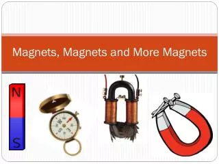
1 Magnets in orthodontics Magnets in orthodontics Prepared by: Prepared by: Dr. Mohammed Alruby Dr. Mohammed Alruby Magnets in Orthodontics Magnets in Orthodontics Dr. Mohammed Alruby Dr. Mohammed Alruby
2 Introduction History Definitions Advantages and disadvantages Types of magnets material Biology of magnets Clinical application of magnets: -Tooth movements -Expansion -Impaction -Open bite -Magnetic functional appliances MAD -Functional orthopedic magnetic appliance FOHA -Magnetic twin block -Magnetic fixed functional appliances -Hemi-facial microsomia -Retainer -Magnetic edgewise bracket -Complex tooth movements Magnets in Orthodontics Magnets in Orthodontics Dr. Mohammed Alruby Dr. Mohammed Alruby
3 Introduction: Magnets from Greek word (Magnesian stone) it is the material or object that produce a magnetic field. This magnetic field is invisible and causes the most property of a magnet. Magnets can be made to attract or repel and therefore to pull or push teeth (Sandler et al 1989) The magnetic force has several advantages than other conventional force: 1-Predictable force level 2-Better directional force 3-No force decay overtime 4-Can exert their force through mucosa and bone 5-Frictional less mechanism 6-Less patients discomfort and less patient’s cooperation There are two types of magnets: 1-Hard magnets (permanent): magnetized without any inducing media and resist to demagnetization 2-Soft magnets: temporary: magnetized due to inducing media and get demagnetized with the absence of induction (electric magnets) History: = ancient people learned about magnetism from lodestone, naturally magnetized pieces or iron ore = the first person writes a book on magnets was William Gilbert in 1600 who realized that the earth was against magnets = serious use of magnets in medicine was reported in early 19 century, a widely recognized use of magnetic force is by orthopedic surgeon to overcome non-union fracture in long bones 1978: Blechman and Kawata introduced Aluminum –nickel – cobalt (Alnico) magnets for orthodontic tooth movements 1984: Muller and Sm Co (samarium- cobalt) magnets for midline diastema closure 1985: Blechman used Sm Co magnets in patients for inter-maxillary orthodontic force 1987: Kawata introduced Sm Co magnetic edgewise orthodontic brackets 1988: Blechman used magnets for complex tooth movement 1989: Vardimon et al introduced functional orthopedic magnetic appliance FOMA for sagittal skeletal correction 1991: Sandler introduce micro-magnets as an alternative solution to fixed retention 1993: Darendeliler introduced magnetic activator devices for sagittal jaw correction MAD 1993: Moss et al describe the use of twin block appliance with magnet incorporated for the treatment of class II division 1 malocclusion 1995: Darendeliler introduced MAD f1995: Darendeliler introduced MAD for open bite correction Definitions: Magnet: material has the ability to attract iron containing alloys and to lie in a north –south direction when freely suspended Hard magnets: magnet that resist to demagnetization in presence of higher magnetic field, at higher temperature and when its size decrease to 1mm or less Soft magnet: magnet has properties that opposite to that of hard magnet, become demagnetized in presence of higher magnetic field, at higher temperature and when its size decrease to 1mm or less Magnets in Orthodontics Magnets in Orthodontics Dr. Mohammed Alruby Dr. Mohammed Alruby
4 Flux density: known as magnetic induction and denoted by B. It is magnetic field strength on the pole face of magnet. Flux density is measured by Hall probe. Its unite are: gauss, tesla, Millitesla. Coercive force: Known as field intensity and denoted HC, it is the strength of the demagnetization field where flux is zero, B=0 Coercivity: the strength of the external field needed to demagnetize the material. Known as the resistance to demagnetization. High coercivity is needed to prevent the magnetization of permanent magnet when they encounter fields produce by other sources Maximum energy production: It is the highest energy that magnet can provide. It is denoted by BHmax. Remanence: it is the flux density in a magnetic material at zero air gap which remain after the removal of the magnetizing field. I = it is denoted by Br. And unites are: Gauss, tesla and Millitesla Isotopic: it is the property that allow crystals of magnet to be aligned in any direction and capable of being magnetized in any direction Anisotropic: It is the property that allows crystals to be aligned in one direction and capable of being magnetized in one direction Anisotropy can be natural or can be induced by hammering, rolling or drawing. Curie point: It is the temperature at which the magnets lose its magnetic properties when subjected to heat At curie point, magnetic domains return to random distribution and as a result magnets lose their properties Magnetic field: It is volume in the medium surrounding the magnet in which its magnetic effects are measurable Magnetic field lines: They are energy lines passing from the north pole of the magnet to the south one in an archial or circular fashion They are more intense at the poles and almost absent at the middle of the magnet Magnetic flux: It is the number of the magnetic lines, passing through a certain area, its unit is weber The flux produced by the magnets causes them to attract or repel other magnets and attract material containing iron The inter-magnetic force has two types: = repulsion: which occurs between two similar poles (N-N or S-S) = attraction: which occurs between two different poles (N-S) Advantages of magnets: 1-Able to produce a measured force continuously over long periods of time, no force decayed over time 2-Less patients discomfort particularly in the initial stage of treatment 3-Force system is frictionless 4-Can be made to attract or repel and therefore to push or pull the teeth Magnets in Orthodontics Magnets in Orthodontics Dr. Mohammed Alruby Dr. Mohammed Alruby
5 5-Force deliver can be directed 6-They can exert their force through mucosa and bone as they does not need to be a direct contact between them 7-Better force and working range control, by maintaining the distance between two magnets better force level can be controlled 8-More biologically acceptable tooth movements 9-Minimum appliance adjustments 10-Less chair side time 11-Minimum patient’s cooperation 12- Removing the need for elastics and springs etc. 13- Magnetic force produces an electro-current (piezoelectric) which will remodel alveolar bone Disadvantages: 1-Corrosion of the magnets can be avoiding by seal it with thin layer of parylene 2-The force produced between any two magnets falls dramatically with distance 3-Bulkiness of magnets 4-Brittle in nature 5-Significant irreversible force is seen if the magnets are heated 6-Difficult to design appropriate size of the magnets as they are very hard and brittle 7-Patients / dentists may be worried by unknown phenomena related to magnetism Types of magnetic materials in dentistry 1-Aluminum-nickel-cobalt (ALNICO) magnet: = since 1953, this type is used widely in dentistry = offer high field strength = have a highest working temperature = they are physically stronger than any other magnetic materials = commercially it is available as rod, bar, ring form = the wide spread use of these types was prevented because of the size, coast, risk of demagnetization magnetic properties were inadequate large flux leakage 2-Rare earth magnets: =rare earth metals are incorporated in the magnets to: -Increase their ability to be magnetized -Get coercivity property - Increase curie temperature = rare earth magnets produce high produce high force relative to their size due to the property of magneto-crystalline anisotropy = when magnets are heated to even modest temperature, they suffer irreversible magnetic loss, as when embedded magnets in acrylic and expose to curing temperature so, there significant amount of flux loss. Advantages: 1-High energy product value 2-Permanent magnets 3-Predictability of amount and direction of force Magnets in Orthodontics Magnets in Orthodontics Dr. Mohammed Alruby Dr. Mohammed Alruby
6 4-Force directly proportion to size and shape Disadvantages: 1-Brittle in nature 2-Low corrosion resistance A-Samarium cobalt magnet (Sm Co): = introduced by Becker in 1970, but the original one developed by Kar / strnat 1960 = available as anisotropic form = better thermal stability = better corrosion resistance = high magnetic field 10 times than ALNICO magnets = corrosion resistance for artificial saliva is good = low resistance to acid attack B-Neodymium-iron-born magnet: NDFeB: = introduced by Robinson 1948 which have a high energy product than SM Co = 240 times susceptible to corrosion than Sm Co magnets = 10 times more powerful than ALNICO magnets Types: Neo 1i: cheaper and sufficient resistance to corrosion Neo 3i: withstand demagnetization at higher temperature but poor resistance to corrosion Neo5i: superior energy production and resistance to demagnetization Advantages: High energy product value Better biocompatibility Strong force attraction Disadvantages: 1-Susceptible to corrosion 2-Risk of destroyed magnetic properties and forces 3-Necessary to coat to prevent cytotoxic product in the oral cavity 3-Pot magnets: = consists of a cylindrical magnet assembly with a concentric nano-magnetic ring into a mid-steel, brass or stainless pot = corrosion is one of the major problem in clinical use of magnets so should be coated by parylene, nickel during clinical use Biological safety of magnets To evaluate biological safety, 3 level of testing are conducted: I: in vitro testing to evaluate toxic and carcinogenic effect II: testing on testing animals III: clinical trails Cell culture studies: = no significant effect on growth rate or type of cell response = no cytotoxic effect on fibroblast like cells = no effect on cell activity either attractive or repulsive field Magnets in Orthodontics Magnets in Orthodontics Dr. Mohammed Alruby Dr. Mohammed Alruby
7 = no significant effect on DNA synthesis, DNA content, cell shape, surface structure or cell number = magnetic bracket produce field influence the oral microbial flora and stimulate the growth of candida albicans. Systemic effect of magnets: 1-No toxic effect by exposing animals to magnetic field strength 2-Did not produce any adverse effect on blood cells 3-No increase in urinary cobalt level at six months’ intervals of magnetic use 4-No significant change in ascorbic acid, calcium and citric acid concentration levels in blood after clinical use of magnets Local effect of magnets: 1-Electromagnetic field clinically enhance the circulation process in fractured bones 2-Reversible epithelial thinning 3-No effect of Sm Co on cellular growth 4-No effect on osteoblast like cells but production of alkaline phosphatase level is increase (double) 5-Orthodontic magnetic brackets affect microbial flora and stimulate the growth of candida albicans 6-Static magnetic field inhibit the osteoblastic activity 7-Static magnetic field reduce the thickness of sebaceous gland and complete disappear after four weeks’ periods 8-Static magnetic field increase proliferation of fibroblasts 9-Static magnetic field cause reversible epithelial thinning Animals studies: in dog mandible: 1-No adverse effects on blood cells 2-No abnormalities of tissue around magnetic infants 3-No change in dental pulp, gingival tissue, periodontal tissue, buccal mucosa, alveolar bone, in the presence of magnetic exposure up to 95 millitesla 4-No abnormal healing or osteoblastic activity 5-No notable difference in cell size, shape or content Other studies: = the pulsed electromagnetic filed and static one enhance the amount of bone formation and hard tissue density at the osteotomy site in guinea pig mandible = increased WBC count in blood = reversible reduction in the number of epithelial cells = the fracture bone unit has no histological change but stronger callus formation between bone Human studies: = implantation of magnets into the molar region at mandible -No significant changes in ascorbic acid, calcium or citric acid concentration -No effect on maxillary buccal mucosal blood flow and pulpal tissue Magnets in Orthodontics Magnets in Orthodontics Dr. Mohammed Alruby Dr. Mohammed Alruby
8 Clinical application of magnets 1-Tooth movements: Muller 1984 suggested that small magnets approximately 5x3x1 could be used to deliver light continuous force to close diastemas without arch wires {simple tooth movements without arch wire} Blechman 1985 use mini-magnets attached to edgewise appliance to move tooth along arch wires Canine retraction: = 1979 Blechman and Smiley: Study using ALNICO magnets on cats, that coated with fast curing acrylic resin to distalize the canine over a period of 9 months to: Produce continuous force and consistent More rapid distalization Less trauma and safe in the oral environment = 1975 Blechman: Using Sm Co magnets in conjunction with edgewise appliance to distalize the canine and found that it is more superior to class II elastics 1987 Kawata et al: Magnets reducing the treatment time, no pain nor discomfort, no periodontal problem, no root resorption, no caries = 1996 Daskalogiar Makis and Mclachlon: They found that canine retracted with a constant force moved more than the control canine with the rate of 2:1 Molar distalization: = Gianelly et al 1988 used repelling magnets for 1st time for molar distalization and recorded rate 3mm in 7 weeks when 2nd molar absent and 0.7 mm / month when second molar are present Design: Modified Nance at 1st and 2nd premolars activated against the maxillary 1st molar to move them distally at rate 3mm / week They found that 80% of space created represented distal movement of 1st molar at rate 0.7mm/month even in the presence of the 2nd molars, result in less anchorage loss Force value is 200gm--225gm at zero gap and 75gm at 1mm air gap About 1mm mesial movement of anchor unit occurs during molar distalization process and this can be reduced by wearing class II elastics of 200gm force during night time Advantages: 1-Better patient tolerance 2-Easy to insert magnets 3-No need for patient cooperation Bondemark et al 1994: Examined repelling magnets versus super-elastics nickel titanium coils used for distal movement of 1st and 2nd molars The result show: 3.2mm distal movement of coil and 2.2mm distal movement of magnets They concluded that: 1-Super-elastics nickel titanium open coil more effective than repelling Sm Co magnets 2-Coil force more constant than magnets forces 3-Coils more comfortable than magnets Magnets in Orthodontics Magnets in Orthodontics Dr. Mohammed Alruby Dr. Mohammed Alruby
9 Diastema closure: Small magnets can be used to deliver light continuous force to close midline diastema without arch wire (Muller1984) Magnets should be fixed to the labial surface of central incisors by indirect bonding, proper position of magnets is very important Advantages: 1-Minimum tooth tipping 2-Not required activation 3-Less chair time 4-Better oral hygiene 5-Magnets can be used after sterilization 6-Position of tooth can be controlled by changing the position of magnets Disadvantages: 1-Difficult positioning of magnets 2-Loss of magnets may occur 3-Fracture of magnets can occur Extrusion of posterior teeth: = one of modalities can be used for correction of deep bite = magnets should be bonded on occlusal surface or on labial surfaces of posterior teeth = tooth movement is rapid and about 2mm / month Intrusion of posterior teeth: = recommended for treatment in anterior open bite cases, using repelling magnets Methods of intrusion: 1-1st permanent molars have magnets of (5mmX5mmX2mm) with 690gm force. Bonded on the occlusal surface with the pole repelling to each other 2-Bonding magnets to the lingual and buccal surface of 1st molars Intrusion of anteriors: In cases of gummy smile where the upper incisors down far below the lip line, attractive magnets are used for intrusion of incisors Steps: 1-Bonding magnets to labial surface of anterior 2-Full covering occlusal splint with bucco-lingual extension containing the magnets To prevent proclination of incisors during intrusion, the labial / buccal acrylic should just touch the labial surface magnets, teeth should be moved one or two at a time in order to minimize anchorage problem 2-expansion: Application of magnets in orthodontic extended to involve orthopedic expansion of the palate and correction of dental cross bites. 1987, Verdimon et al used repulsive magnetic force for maxillary expansion versus mechanical expansion with different force threshold (on four juvenile Macaca monkey) Study design: One received conventional type jackscrew maxillary plate bonded to posterior teeth with high force magnitude Second: tooth born repulsive magnets having a low force of 220gm Magnets in Orthodontics Magnets in Orthodontics Dr. Mohammed Alruby Dr. Mohammed Alruby
10 Third: especially designed palatal acrylic appliance directly to palatal shelves with rare earth magnets with low force Fourth: as control Conclusion: 1-Magnetic appliances deliver force in superior lateral direction which dissipated in the zygomatico-frontal suture 2-Increased overjet due to widening of transverse and incisive sutures 3-High affect was in molars area 4-Recommend to correct collapsed maxillary and pre-maxillary sutures Advantages of magnetic over conventional type: 1-Produce controlled force over predictable range. 2-Low expansion as compared to rapid one 3-Force is more physiologic so it avoids the complications of rotation of maxilla ** Vardinon et al 1989: Concluded that, stability of magnets palatal expansion was more than rapid expansion ** Darendeliler 1994: Found slow expansion magnets more effective specially at an early age (50% skeletal effect can be obtained) Magnetic field also enhance tooth movement so rapid rate of expansion 3-Impaction: The use magnets reported firstly by McCord 1984 to extrude teeth, by extruding the roots a sub- gingivally fracture incisors by Sm Co magnets, one fixed to the embedded root and the other embedded in the removable partial denture = 1989 Sandler et al: 1st use magnets to guide in the eruption of an impacted canine = 1991 Vardimon et al: utilized technique in the guided eruption of impacted teeth, they used magnetic bracket Nd2 Fe14 B bonded to an impacted teeth and an intra-oral magnets linked to a Hawley retainer = 1994 Darendeliler and Friedli, used a combination of removable and fixed type attraction for impacted canine, in which the fixed part fixed to sectional arch = 1997 Bondemark et al, used Nd Fe B to extrude, the remaining roots the roots were extruded 2- 3mm with force range 50 – 30 during treatment period of 9-11 weeks 4-Open bite: Dellinger 1986 was the 1st use magnets in the correction of anterior open bite, he introduced the active vertical corrector AVC Mode of action: Magnetic field produce micro-current flow in the periodontium which increase the cellular activity and being rapid tooth movement Intrusion of posterior teeth upper and lower Upward and forward autorotation of the mandible Closure of anterior open bite, followed by reduction of anterior facial height Appliance design: Removable or fixed appliance with acrylic bite block with Sm Co magnets to intrude the molars One or two magnets / quadrant can be used depending on the force requirement Force produced by the appliance is 600—700gm at zero air gap 12 hours / day is recommended at least but 20 hours is better correction can occur within 4 -9 months of treatment Magnets in Orthodontics Magnets in Orthodontics Dr. Mohammed Alruby Dr. Mohammed Alruby
11 == Dellinger stated that, problems formerly treated surgically can now be treated by AVC successfully Other studies: 1-Woods and Nanda 1988: using bite blocks and they found that there are similar results with both magnetic and non-magnetic bite 2-Melson et al 1991 found that using magnetic bite blocks caused unerupted teeth to show more root formation with an inverted Hertwig root sheath 3-Kuster and Ingervall 1992 reported that beneficial effects of the treatment tend to relapse 4-Darendeliler et al 1995: correct open bite by magnetic activator device IV MAD IV 5-Magnetic functional appliances: magnetic activator device: Introduced by Darendeliler in 1993, there are four varieties for magnetic activator device: -MAD I: for correction of lateral mandibular displacement -MAD II: for correction of class II -MAD III: for correction of class III -MAD IV: for correction of open bite MAD II: magnetic activator device II: = introduced by Darendeliler in 1993, the magnets impeded in acrylic portion of upper and lower = if the appliance could displace the condyle downward and forward away from the posterior part of glenoid fossa, stimulate the condyle growth that lead to increase the length of mandible and improvement in the facial profile Design: = upper and lower removable acrylic plate carrying magnets in both buccal segments = each one has lingual acrylic portion with C-clasp in 1st molars, labial bow, two Sm Co magnets and occlusal bite plane to avoid cuspal interferences = magnetic size 4 x 4 x 6 x 1 = magnets are placed in buccal aspect of the appliance with 30 degree angulation to the buccal surface that produce oblique force to correct Class II =in class II with normal mandibular growth (with average FMA) magnets are placed distal to the lower 1st premolar = in class II deep bite (low FMA), magnets are placed an attractive force between them to produce extrusion of molars and premolars = in class II open bite (high FMA), two repelling magnets can be used posteriorly, produce molar and premolar extrusion with some distal movement of upper arch = force value is 300 -350gm / side producing total 600 -700gm = the distance between two magnets anterior- posterior should not be more than 7mm, if it is more than 7mm the magnetic force will produce less than 35gm of force and not adequate to guide the mandible in anterior position = wax bite registration is similar to that for conventional functional appliances = position and angulation of magnets depend on: 1-Center of resistance of upper arch 2-Maxillary and mandibular plane inclination 3-Facial height ** if vectors of attracting forces pass anterior to the center of resistance it will produce downward and backward rotation of maxilla ** if vectors of attracting forces combined with repelling force vector pass posteriorly to the center of resistance, will produce downward and backward rotation of maxilla and upward and forward Magnets in Orthodontics Magnets in Orthodontics Dr. Mohammed Alruby Dr. Mohammed Alruby
12 rotation of mandible which is force vector pass posteriorly to the center of resistance, will produce downward and backward rotation of maxilla and upward and forward rotation of mandible which is favorable in high angle cases MAD III: magnetic activator device III: = introduced by, Darendeliler, Chiarini and Goho 1993 = produced light maxillary expansion forces combined with functional orthopedic component for early correction of class III malocclusion Effect of MAD III: 1-Stimulation of forward maxillary growth 2-Maxillary expansion 3-Restriction in forward mandibular growth Design: = the upper appliance canine to buccal magnets which are attractive in nature, two repelling magnets for palatal expansion = the lower appliance carries two buccal magnets which are placed more bucally and anteriorly than the upper buccal magnets so that their attractiveness force would not interfere with the repulsive forces of mid palatal magnets = the upper and lower magnets have the tendency to move toward a fully centered contact creating forward force for maxilla and backward force for mandible = expansion force produced by magnets is 800gm, 500gm for palatal magnets and 300gm for buccal magnets, sagittal force produced is 600gm from each magnets Advantages: 1-Increase the length of maxilla 2-Restrict mandibular growth 3-No problem in speech, swallowing and chewing 4-Magnets can be relocating to orient the inter-maxillary force Darendeliler et al 1993, studied two case treated successfully by MAD III, one case combined with magnetic expander device and the other with Delaire mask Moss et al 1993: described the use of the twin block appliance with magnets incorporated in treatment of class II division 1 malocclusion, noted that incorporate magnets into the appliance decrease the time taken to produce the sagittal changes and increase the soft tissue changes compared to those appliances without magnets MAD IV: magnetic activator IV: = introduced by Darendeliler, Yuksel, and Meral 1995 = the appliance consists of removable upper and lower plates; each plates consists of three cylindrical magnets coated with stainless steel = upper and lower plates contain anterior attracting magnets and posterior repelling magnets Types: a-Used where posterior intrusion and mandibular autorotation is required (gummy smile) b-When additional extrusive effect on anterior part of maxillary is necessary c-If only extrusion of anterior teeth is required All these types caused: -Reduction in anterior height -Increase incisors -Sagittal growth modification Magnets in Orthodontics Magnets in Orthodontics Dr. Mohammed Alruby Dr. Mohammed Alruby
13 6-Functional orthopedic magnetic appliance: FOMA: = introduced by Vardimon et al 1989, which has two criteria: 1-Has intrusive force built in 2-Force is continuous in nature Types: FOMA II: for class II skeletal correction FOMA III: for class III skeletal correction A-Functional orthopedic magnetic appliance II: FOMA II: Design: = the appliance consists of upper and lower plates = the magnets are incorporated in the plates are attractive in nature = maxillary magnets are a head of mandibular magnets; thus the resulting attraction maintain the lower jaw in an advanced posture = upper and lower magnets hold the lower firmly in an advanced sagittal posture = reactivation of the appliance can be done by relocating the upper magnets forward The increase in mandibular length is due to: 1-Posterior superior endochondral growth 2-Bone remodeling at neck of condyle Results: -Significant increase in mandibular length -Minimum incisors proclination B-Functional orthopedic magnetic appliance III: FOMA III: Introduced by Vardimon et al 1990, it is an intra-oral appliance for correction of class III malocclusion, that inhibit midface sagittal deficiency with or without excess Design: =upper and lower plates with magnetic arranged in an alternative force Upper magnets are posterior to lower arch and lower magnets is posterior to plate near the lingual surfaces of incisors = horizontal force produced by the magnets is 98gm and vertical force produced by magnets is 371gm = reactivation of the appliance is done by posterior re-positioning of the upper magnets in every 3-4 weeks 7-Magnetic Twin block: Moss et al 1993 described twin block appliance with magnets incorporated for class II division 1 treatment. He reported that, with magnets sagittal correction is faster and also enhanced soft tissue changes 8-Magnetic fixed functional appliance: Introduced at 1989 by Katara, Nanda, Burstone, for correction of class II division 1 Functions: 1-Improve the angle of convexity 2-Intrude teeth 3-Auto-rotate mandible in upward and forward direction 4-Increase in the length of the mandible Magnets in Orthodontics Magnets in Orthodontics Dr. Mohammed Alruby Dr. Mohammed Alruby
14 Design: = upper and lower acrylic splint that were bonded on the occlusal half of the 1st permanent molar, deciduous molars and canines = magnets encase in st st cases and embedded into the upper and lower acrylic splint in repelling mode = 0.028 wire embedded in the acrylic and rest on the lingual surfaces of incisors and bonded so the intrusive force will transmit to the entire arch Bite registration: Mandible at rest position and 7 -8mm opening at 1st molar region Main disadvantages: Creation of temporary cross bite by shearing force of repelling magnets ** Karata et al 1989 reported that after 4 months’ active treatment, a significant increase in mandibular length and decrease in the mandibular plane angle 9-Hemifacial Microsomia: Chate in 1995, presented a new magnets Sm Co embedded in unilateral acrylic block (PUMA) (propellant unilateral magnetic appliance) to stimulate the autogenous costo-chondral graft = this appliance involved unilateral blocker of acrylic in separate upper and lower removable with cylindrical gold plated magnets, that incorporated with their long axis in repelling mode perpendicular to the blocks interface, and separated by 3mm 10-Retainers: Dahle and Zachrhrison 1991, use fixed retainer to stabilize the anterior spacing 1995, micro-magnetic retainer suggested by Springate and Sabdler to retain central incisors Advantages of magnetic retainers: 1-Oral hygiene can be maintained as flossing is not prevented 2-No wires or ledges close to the gingival margins 3-Teeth not splinted together 11-Magnetic Edgewise brackets: = kawata et al 1987, introduced magnetized bracket and claimed that magnetic force applied to move the teeth, produced less stress than the conventional method of force application with the help of springs, coils and elastics Bracket design: = Sm Co magnets of suitable size 6 x 2 x 1 m and 3 x 2 x 1 = standard edgewise design with 0.018 x 0.025 slot = chromium coating over the magnets = soldering of magnets to the bracket with nickel (nickel allows the bracket to be soldered to the surface below 500 degree) = soldering a mesh base onto the magnetic bracket so that bracket can be directly attached to the tooth surface Bracket size: Upper: M D: 7mm, Occ Gn: 3.5mm, L L: 3mm Lower: M D: 4mm, Occ Gn: 3.5mm, L L: 3mmorce These bracket produce mesiodistal magnetic of 50 – 250gm and are mostly used for canine retraction Magnets in Orthodontics Magnets in Orthodontics Dr. Mohammed Alruby Dr. Mohammed Alruby
15 N: B: Magnetic brackets are however discarded because of complexity of lab preparation and difficulty to obtain proper dimension of magnets necessary for optimum force level 12-Complex tooth movement along arch wire: Blechman in 1985 concluded that, permanent magnets can be used for inter-maxillary and intra- maxillary mechanics in attraction or repulsion to move teeth along arch wire A-Class II mechanics: = magnets assembly is attached to an upper sectional wire which slides freely through the occlusal upper molar tube, ligated mesially to upper canine bracket. = lower magnets are attached to a similar sectional arch passing through the occlusal tube of the lower molar = upper and lower magnetic pols face each other in an attraction mode in order to generate the force to move the upper canine distally along the base arch wire and lower buccal segment mesially along the base wire B-Intra-maxillary magnetic force to move canine distally: magnets can be positioned on separate arch wires in order to effect tooth movement C-Class II mechanics in class II non extraction cases Magnets in Orthodontics Magnets in Orthodontics Dr. Mohammed Alruby Dr. Mohammed Alruby