Download

1 / 8
0 likes | 3 Views
Notes in Orthodontic Cephalometry

E N D
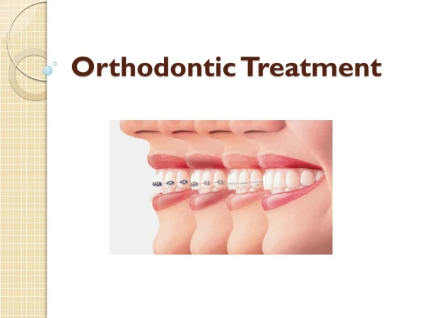
1 Notes in orthodontic cephalometry Notes in orthodontic cephalometry Prepared by: Prepared by: Dr Mohammed Alruby Dr Mohammed Alruby اثيدح مهلقا سانلا قدصا Notes in Orthodontic Cephalometry Notes in Orthodontic Cephalometry Dr. Mohammed Alruby Dr. Mohammed Alruby
2 History of X-ray: 1895: 8/11/1895: Conard Reontegn discover X-Ray 1877: Conard Reontegn take an X-ray film on his wife hand 1922: lateral head film by Perciri 1922: first x-ray on skull – standard lateral film by Pacini and Carrera 1922: Atkinson used lateral film to locate Key ridge 1928: Macleven used profile photograph to detect changes in profile 1928: Deway and Raiser: immobilize the head in head clamps and oriented the FH by right angle and use distance 3 feet 1931: Broadbent and Hofrath developed standard cephalometric technique using special holder known as cephalostat which permit careful growth assessment x-rays: form of electromagnetic radiation that penetrate the body which allow radiologist to produce pictures of internal structure. Have wave length ranging from 0.01 to 10 nanometer Have frequency ranging from 30 pet hertz to 30 hexa hertz Uses of cephalometry: 1-Diagnosis: assessment Ap and Vertical, skeletal pattern, incisors position, and soft tissue 2-Treatment planning 3-Monitoring the treatment 4-Research and audit Reference lines: == horizontal lines: 1-FH 2-Max plane 3-Mand plane 4-Occlusal plane 5-Sn 6-True horizontal: Sallow and Tallegram 1971: 7 degree from Sn 7-Decoster line supra- orbital plane == vertical lines: 1-N-Pog: facial plane: Downs 2-A-Pog: Willians 1969 3-Zero meridian: Nasion perpendicular to FH 4-Pterygoid vertical: line perpendicular to FH and posterior margin of pterygoid palatine fossa 5-H line 6-E line Protection from radiation x-ray is a form of electromagnetic radiation that can be cause biologic changes to a living organisms by ionizing the atom in the tissue they radiate: 1-Utilization high speed film and intensifying screen to reduce the dose of radiation 2-Filtration of secondary radiation and scattered that produced by lower energy x-ray photons 3-Collimation by diagram lead to optimum beam size 4-Patient wear lead apron in order to absorb scattered radiation 5-Operator should stand six feet behind the tabs head. Notes in Orthodontic Cephalometry Notes in Orthodontic Cephalometry Dr. Mohammed Alruby Dr. Mohammed Alruby
3 Posterior anterior cephalometry: For face breadth and a symmetry =Useful information concerning over all morphology, shape and size of skull =Useful for patient seeking to orthognathic surgery for facial a symmetry Limitation: Precise measurements of detail are likely to be misleading Tilted head ----- change in parameter ------- angular or linear Submento vertex: basilar cephalometry =Radiograph projection showing: base of skull, position of mandibular condyle, zygomatic arches =Show pathology of base of skull and sphenoid sinus =Central rays directed to the mid-sagittal plane at point mid-way between the angle of mandible which is also approximately the point mid-way between external auditory meatus =are covered; occiput, foramen oval, foramen magnum, sphenoid sinus Source of error in lateral cephalometry: Validity: accuracy: is the extent to which in absence of measurement error (Hauston 1983) Reproducibility: closeness of successive measurements of the same object ( Hauston 1983) Reliability: both validity and reproducibility 1-Protection errors; a-Magnification: proportional enlargement of all parts structure depend on the distance from object- film – x-ray source b-Distortion: lacke of exact reproduction of structure in term of proportion Different parts of structure are not increase proportionally In lateral film the only part that not distorted those in midline, but bilateral structure is very important to overcome this problem c-Blurring: lack of sharpness of the radiograph Factors affect: Movement of patient Time of exposure Secondary radiation Distance film – object – source 2-Errors within the measuring system: Error for recording procedure has two components a-Identification of tracing points, by device used b-Error in digitizing system 3-Errors in landmark identification: Considered as the major source of cephalometric error (Bjork 1947), Depend on: a-Quality of radiographic image ---- blurring Contrast; is the magnitude of the optical density Differences between a structure and its surroundings Contrast determined by: tissue being examined, receptors, level of K.V used b-Precision of landmark definition, location and reproducibility c-Operator and registration procedure. Errors in growth prediction: 1-Wide range of morphologic differences 2-Varying rates and direction during growth periods 3-Varying influences of modifying environmental factors 4-Variation in trimming of the different areas of active growth 5-Lack of correlation between the size of facial structure at an early age and ultimate size. Notes in Orthodontic Cephalometry Notes in Orthodontic Cephalometry Dr. Mohammed Alruby Dr. Mohammed Alruby
4 6-Cephalometric head film taken at different times and by different techniques so it difficult to be reproduced with high degree even if head in cephalostate 7-Double image of any bilateral structure due to minor fault head position 8-Difference in film contrast and density due to lack of quality control 9-Anatomical or constructed landmark are not easily identified 10-Cephalometric are 2D for 3D object, so we recommend computerized 3D technology in radiographic usage Effective dose in radiograph: Upper occlusal ---- Usv Panorama ----- 3 –24 Usv Lateral ceph. ----- > 6 Usv Con beam Ct ----- 50 –500 Usv Requirement of cephalometric landmark: 1-Easy to seen, uniform in outline 2-Should has a significant relation to the information 3-Able to statistically analyzed 4-Have significant relationship to the growth vector of a specific area. Reliability of cephalometric landmark: cephalometric landmarks are subjected to variation due to growth changes of individual bones over which these bones are down like Sn line, is not considered as fixed reference as this line pass through different bone (nasal, ethmoidal, sphenoid) these area was affected by growth which affect cephalometric finding =Bolton, Basion points are functionally related to the cerebellum and not a part of cranial base, so they are variable. =Gran 1961, pointed out that, there are no fixed points in the skull but most landmark are variable, however lesser changes occur in landmark near to cranial base. Variability depend on: Age, maturation rate, sex, ethnic background = the more variable landmarks are Bolton, basion, porion, Gonion, orbital, ANS, Apoint, PNS, Bpoint = over knowledge about anatomy, biology, are very important to overcome the problem of variability and distortion of measuring point. Reliability of lower border of mandible: Lower border depends on location of Gonion which migrate backward and vertically during growth = tangent to lower border of mandible not reliable but, Go—Me and Go – Gnth are good Occlusal planes: 1-Line joining midpoint of overlapping buccal cusps of 1st molars with point bisect the over bite of incisors (Downs 1943, Steiner 1943) 2-Bisect the buccal cusps of molars and premolars or buccal cusps of deciduous molars (Ricketts 1960, 1961) (Jacobson 1975—wits analysis) 3-Line join midsection of molar cusps and tip of upper incisors (Bjork 1947) Mandibular planes: 1-Tangent to lower border of mandible (Tweed 1947, Wylie 1947, Ricketts 1960) 2-Line from Gonion to Gnathion Steiner 1953 3-Line from Gonion to Menton (Downs 1948, Milles 1070) Authors and analysis: 1-Upper gonial angle + lower gonial angle ------ Jaraback 1972 2-H angle , H line ------------ Holdaway 1983 Notes in Orthodontic Cephalometry Notes in Orthodontic Cephalometry Dr. Mohammed Alruby Dr. Mohammed Alruby
5 3-Holdaway ratio: Li – NB / pog – NB -----Holdaway 4-Cc point, Xi point, PM point ------- Ricketts 1957, 1960 5-Facial angle ----- Ricketts 6-DC: point at which center of condyle as nasion basion line cross it ---------Ricketts 7-Sassoni analysis ------- Sassoni 1955 8-U1 to Na linear and angular ----------Steiner 1953, 1959 9-L1 to NB linear and angular----------- *** ** ** 10-L1 to Go ---Gnth -----------** ** *** 11-U6 to NA -------------** ** ** 12-L6 to NB --------------** *** ** 13-SND angle ( D: center of symphysis) -----** ** ** 14-Tweed analysis -------- Tweed 1946, 1953 15-Downs analysis: ---------- William Downs 1948, 1956 16-Angle of convexity ------------------------ Downs 17-Y axis --- ----------------------------------- Downs 18-Articular angle S Pr G ----------Bjork 19-Saddle angle N S Ar ----------Bjork 20-S N line ---------------- Bjork 21-Macnamara anlysis -------------James Macnamara 1984 Profile angles: 1-Nasofacial angle: 20—35 2-Nasomental angle: 120—132 3-Mentocervical angle: 110—120 4-Submental neck angle: 126 in males, 121 in females 5-Nasolabial angle: 90—110 Subtelny profile analysis: 1-Skeletal profile: N A Pog decreased by age 2-Soft tissue profile: N* Sn Pog* constant 3-Full soft tissue profile: N* Prn* pog* increased by age Bjork analysis: 1-Angle at N --- SNA 2-Saddle angle ---- S N Ar 3-Articular angle --- S Ar Go 4-Jaw angle ----- Ar Go Me Wits analysis: Alexander Jacobson 1975, 1976 In Witwatersrand in south Africa Sample of 21 adult males and 25 adult females with good occlusion Line perpendicular from A point and from B point to functional occlusal plane and measure the distance between the two points Norm.: 1mm males -+1.9 and 0 mm in females -+1,7 Ballard conversion: = Cliford Ballard describe simple method for assessing A.P jaw relationship using axial inclination of incisors (Ballard 1951) = trace on a separate piece of tracing paper outline line of maxilla, symphysis = mark rotation points of incisors one third of root length away from root apex = rotate the new position around this point ------- normal value Notes in Orthodontic Cephalometry Notes in Orthodontic Cephalometry Dr. Mohammed Alruby Dr. Mohammed Alruby
6 = the residual over jet reflect the underlying skeletal pattern ** validity of Ballard conversion depend on: 1-The incisors bear a constant relationship to the jaw position 2-There is an average inclination of the incisors to the dental bases 3-The incisors will be always tip around defined fulcrum (Houston 1975, Akpabis 1979) Assessment of skeletal relationship by Ballard method: A method advised by Ballard 1948 same as residual over jet method = outline upper and lower incisors teeth by tracing it on lateral cephalogram = long axis is drowning from tip of crown to root apex = pivot point is marked on the long axis one third from the root length from the apex = upper incisors are then redrawn by new outline and new long axis but with an ideal angulation to the maxillary plane but passing through the same pivot point ------ U1 to Mx plane – 108 degrees = also was done to the lower with normal angulation to the upper incisors --- U1 to L1 --- 135 degrees Then the remaining over jet is measured to indicate the skeletal type either I or II or III. Mac Namara analysis: By James MacNamara 1984 Use FH plane as horizontal references and constructed a perpendicular line through Nasion to provide vertical reference 1-Maxilla to cranial base: A point -------- Nasion perpendicular 2-Mandible to cranial base: B point --------Nasion perpendicular 3-Maxillary length: condylon to A point 4-Mandibular overall length: condylon to Gnathion 5-Lower anterior facial height LAFH: ANS to Menton 6-U1 to Maxilla: line from facial surface of U1 to line through A point parallel to nasion perpendicular 7-L1 to mandible: from facial surface of L1 to line drawn through A—PogDowns Down’s analysis Williams Down: one of the 1st propose cephalometric analysis (1948, 1952, 1956) Study on 20 Caucasian boys and girls, from 12 to 17 years of age with excellent occlusion and facial harmony Eastman analysis This analysis further developed by Richard Mills 1982 which use the cephalometric of 250 individuals in Eastman hospital in London. Skeletal analysis: SNA, ANB correction, SNB, MMPA, FMPA, SN –Mx plane Dental analysis: U1, L1 –to SN, U1 to Max plane, L1 to Mand. Plane Eastman correction: Richard Mills 1970 Variation in the position of Nasion can alter the SNA value, so the more anterior or superior position of N------------ decrease the SNA value the more posterior or inferior position of N ----------- increase the SNA value so alteration in SNA alter ANB angle Mills introduce correction for erroneous value of SNA as: = for every degree of SNA greater than 81 degrees subtract 0.5 degrees from original ANB angle = for every degree of SNA lesser than 81 degrees added 0.5 to original ANB This can calculate but in case of SN----- Max p should be 8 degrees -+3 X = measured SNA – average SNA / 2 as X is ANB measured Notes in Orthodontic Cephalometry Notes in Orthodontic Cephalometry Dr. Mohammed Alruby Dr. Mohammed Alruby
7 Holdaway 1983 H line: tangent to chin and upper lip, normally lower lip 1---to 2mm to this line H angle: H line with NB line skeletal 10 degrees or 7 –to 14 degrees Jarabak analysis Saddle angle: N S Ar Articular angle: S Ar Go Gonial angle: Ar Go Gnth Upper gonial: Ar Go N Lower gonial: N Go Me Superimposition Color code for tracing Suggested by American board of orthodontics 1990 as follow: Pre-treatment: black Progress: blue End of treatment: red Retention: green 1-Cranial base: At birth: inter-sphenoid, inter-ethmoid synchondrosis are closed by age at 7 years, the only one still work is sphenooccipital synchondrosis (knott 1971) So: there is a little change in anteroposterior direction at ethmoidal portion at anterior cranial base a-Broadbent triangle: N S Bo and its registration at R point (Broadbent 1931) b-S N line Steiner c-S N line and registration at S point (American board of orthodontic 1990) But there are some changes at the areas of cranial base specially growth at synchondrosis (Knott 1971) Most of changes in position of nasion are due to enlargement of frontal sinus and consequently upward and downward migration of frontonasal suture which result in error in superimposition (Nellson 1960, Knott 1971) Sella turcica also undergoes eccentric remodeling during adolescent age d-Basion horizontal: at the level of foramen magnum and parallel to FH, by this method Basion is used as reference point (Coben 1955, 1986) e-Basion nasion plane (Ricketts et al 1979) superimposition at Basion nasion line with facial axis (pt----- Gnth) N: B: pt. pterygoid point ia anatomical point represent radiolucent foramen rotundum N:B: the position of Basion is influenced by remodeling process at the surface of clavius and at anterior border of foramen magnum f-Anterior wall of Sella turcica g-Contour of cribriform plate of ethmoid cells (lamina cribrosa) h-Tuberculum system in the ethmoid cells i-Median border of orbital roof j-Plane of sphenoid bone (planum sphenoidal) k-Decoster line 2-Maxillary superimposition: a-Palatal plane ------ Morree b-Palatal plane: registration at ANS (Broadbent 1973, Ricketts 1960, Macnamara 1981) c-Nasal floor and registed at anterior surface of maxilla ---- Down 1948 d-Palatal plane and registed at pterygomaxillary fissure Ptm (Morree 1959, Coben 1986) N: B: ptm; pterygomaxillary fissure: oval shape between the posterior surface of maxilla and anterior margin of sphenoid bone e-Outline of infratemporal fossa and posterior portion of hard palate (Riedel 1974) Notes in Orthodontic Cephalometry Notes in Orthodontic Cephalometry Dr. Mohammed Alruby Dr. Mohammed Alruby
8 f-Structural superimposition on the anterior surface of zygomatic process of maxilla (Bjork and Skieller 1976) g-Implant (Bjork and Skeiller) 3-Mandibular superimposition: a-Anterior contour of chin b-Border of symphysis c-Inner contour of cortical plate at the inferior border of symphysis d-Third molar tooth buds before root formation e-Contour of mandibular canal Structures used to assess mandibular rotation 1-Symphysis with cortical bone 2-Inferior and posterior contour of mandible 3-Point articulare 4-Anterior contour of the ramus 5-Mandibular canal 6-Third molar buds before root formation 7-First molar 8-S N line 9-Most labially positioned lower incisors Dentoalveolar height = Upper anterior Dentoalveolar height UADAH: line from tip of U1 perpendicular to palatal plane = Upper posterior dentoalveolar height UPDAH: line from tip of U6 mesiobuccal cusp perpendicular to palatal plane = Lower anterior dentoalveolar height: LADAH: line from tip of U1 perpendicular to the mandibular plane = Lower posterior dentoalveolar height: LPDAH: line from tip of L6 mesiobuccal cusp perpendicular to the mandibular plane Cephalometric criteria for deep bite cases 1-Abnormal inclination of upper and lower incisors 2-Excessive over bite 3-The four planes of the face are in horizontal direction or nearly parallel to each other (cranial base line, occlusal plane, mandibular plane, maxillary plane) 4-Small cranial base angle 5-Small gonial angle 6-Small XI angle 7-Large mandibular arc angle 8-Anterior upper facial height within normal range 9-Anterior lower facial height is reduced than normal 10-The facial breadth is nearly equal to the anterior total facial height, anterior total facial height is nearly equal to the posterior facial height, which give square face 11-Small S N ----- Mp angle 12-Small FMPA 13-Posterior maxillary dentoalveolar height is decreased 14-Posterior mandibular dentoalveolar height is decreased 15-More wide symphysis 16-Upper anterior dentoalveolar height may be: a-Normal: so the LADAH increased b-Increased: gummy smile: so the LADAH may be normal Notes in Orthodontic Cephalometry Notes in Orthodontic Cephalometry Dr. Mohammed Alruby Dr. Mohammed Alruby