Download
1 / 7
70 likes | 75 Views
Relationships between Circulating and Intraprostatic Sex Steroid Hormone Concentrations - Muhannad Hafi MD

E N D
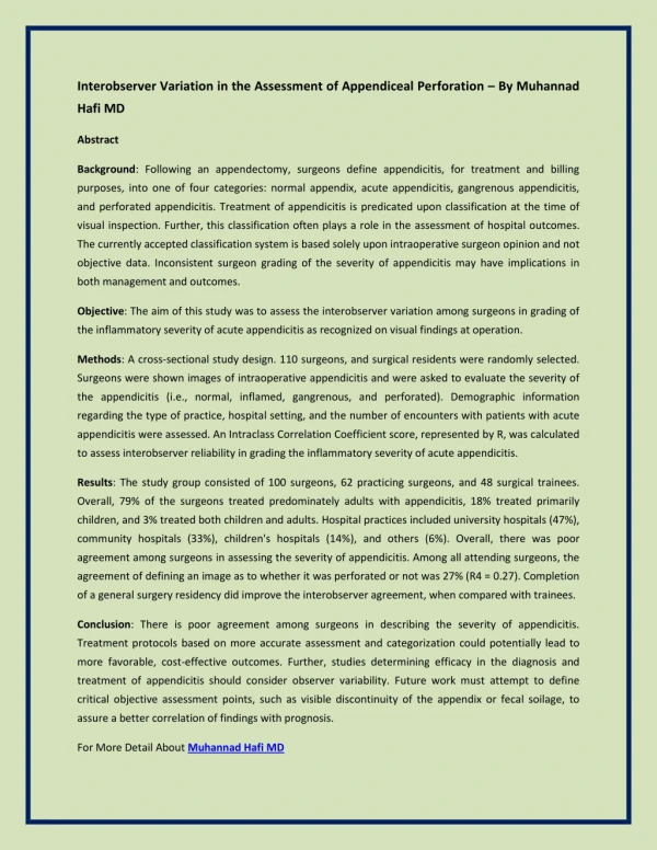
Abstract Background: Sex hormones have been implicated in prostate carcinogenesis, yet epidemiologic studies have not provided substantiating evidence. We tested the hypothesis that circulating concentrations of sex steroid hormones reflect intraprostatic concentrations using serum and adjacent microscopically verified benign prostate tissue from prostate cancer cases. Methods: Incident localized prostate cancer cases scheduled for surgery were invited to participate. Consented participants completed surveys, and provided resected tissues and blood. Histologic assessment of the ends of fresh frozen tissue confirmed adjacent microscopically verified benign pathology. Sex steroid hormones in sera and tissues were extracted, chromatographically separated, and then quantitated by radioimmunoassays. Linear regression was used to account for variations in intraprostatic hormone concentrations by age, body mass index, race, and study site, and subsequently to assess relationships with serum hormone concentrations. Gleason score (from adjacent tumor tissue), race, and age were assessed as potential effect modifiers. Cancer Epidemiology, Biomarkers & Prevention http://cebp.aacrjournals.org/content/26/11/1660
Results: Circulating sex steroid hormone concentrations had low-to-moderate correlations with, and explained small proportions of variations in, intraprostatic sex steroid hormone concentrations. Androstane-3α,17β-diol glucuronide (3α-diol G) explained the highest variance of tissue concentrations of 3α-diol G (linear regression r2 = 0.21), followed by serum testosterone and tissue dihydrotestosterone (r2 = 0.10), and then serum estrone and tissue estrone (r2 = 0.09). There was no effect modification by Gleason score, race, or age. Conclusions: Circulating concentrations of sex steroid hormones are poor surrogate measures of the intraprostatic hormonal milieu. Impact: The high exposure misclassification provided by circulating sex steroid hormone concentrations for intraprostatic levels may partly explain the lack of any consistent association of circulating hormones with prostate cancer risk. Cancer Epidemiol Biomarkers Prev; 26(11); 1660–6. ©2017 AACR. http://cebp.aacrjournals.org/content/26/11/1660
Introduction Prostate cancer has long been hypothesized to have a hormonal pathogenesis. Endogenous sex steroid hormones, particularly androgens, are undoubtedly essential for normal physiological development, maintenance, and function of the prostate gland. Prepubertally castrated men and male pseudo-hermaphrodites with deficient 5α-reductase type II have a mal-developed male phenotype, including a small and immature prostate gland (1, 2). The Nobel Prize–winning studies by Huggins and Hodges in 1941 reported that castration and injection of estrogen cause temporary regression of metastatic prostate cancer, implicating androgenic action in prostate cancer progression (3). This led to development of androgen deprivation therapy, which remains the mainstay therapy for men with advanced prostate cancer. Androgen signaling also functions in cell proliferation, differentiation, and apoptosis, and evidence from basic science indicates that androgens, and possibly estrogens, are critically important for prostate carcinogenesis (4–6). Despite this evidence that implicates sex steroid hormones in prostate cancer pathogenesis, epidemiologic studies that have assessed prediagnostic circulating hormone concentrations have not found any consistent association with subsequent prostate cancer risk (7–9). There are various explanations for why a true association may have been missed, including interassay variability, lack of assay standardization, use of a single peripheral blood measurement typically at middle age or later, and case heterogeneity with inclusion of a variable proportion of indolent disease. http://cebp.aacrjournals.org/content/26/11/1660
Testosterone (T) and the more potent metabolite, dihydrotestosterone (DHT), bind the androgen receptor within the prostate eliciting gene expression profiles and biological effects that maintain prostate function. T is predominantly produced by the testes and released into the circulation. DHT, however, is primarily produced within the prostate gland, thus circulating DHT precursors [T, androstenedione (A)] and metabolites [5α-androstane-3α,17β-diol glucuronide (3α-diol G)] have traditionally been assessed as proxies. The validity of these proxies has not been tested. Therefore, we set out to test the hypothesis that circulating sex steroid hormone concentrations are valid proxies of intraprostatic concentrations using a large set of blood samples paired with microscopically verified benign tissue samples adjacent to prostate cancers. http://cebp.aacrjournals.org/content/26/11/1660
Materials and Methods Study population Patients were enrolled in the study between January 2000 and April 2004 at five locations: George Washington University Medical Center (Washington, DC), University of California at San Francisco (San Francisco, CA), Doctor's Community Hospital (Lanham-Seabrook, MD), Washington Hospital Center (Washington, DC), and INOVA Fairfax Hospital (Falls Church, VA), the latter three of which were primarily coordinated by the staff at George Washington University Medical Center. Study subject eligibility included: 18 years of age or older; scheduled for radical prostatectomy; and newly diagnosed with localized prostate cancer. Patients provided written informed consent to be part of the study. Prior to surgery, study patients had standard anthropometric measures taken and were administered a questionnaire to confirm that they were fasting and had not taken any hormones (e.g., DHEA) or substances that could potentially affect hormone concentrations (e.g., finasteride) in the preceding 24 hours. Study subjects also provided 30 mL of blood, which were processed within 4 hours into aliquots of serum, plasma, buffy coat, and red cells, and subsequently stored for long-term storage at −70°C. http://cebp.aacrjournals.org/content/26/11/1660
During surgery and immediately after the prostate had been resected, the pathologist conducted a sterile dissection of macroscopically benign tissue to obtain a maximum of three peripheral and three periurethral tissue samples, each weighing 200–400 mg. Each macroscopically benign tissue sample had the ends trimmed and placed in formalin for hematoxylin and eosin slide preparation for morphologic and histologic evaluation (by IAS) to ensure that these trimmed ends of the tissue sample for hormone analysis were free of cancer. The main central piece of the tissue sample was placed in a prelabeledcryovial, flash frozen in liquid nitrogen, and stored at −70°C. All collected samples remained available for diagnosis until the surgical pathology report was deemed to be complete. After surgery and histologic assessment of the remainder of the prostate, medical records and pathology review forms were abstracted. A 30-minute telephone questionnaire was administered to study subjects approximately 6 weeks after surgery to elicit information on personal characteristics, medical history, family history of cancer, medication use, and lifestyle exposures. This study was conducted in accordance with recognized ethical guidelines (e.g., Declaration of Helsinki, CIOMS, Belmont Report, U.S. Common Rule) and was preapproved by the required institutional review boards. http://cebp.aacrjournals.org/content/26/11/1660
During surgery and immediately after the prostate had been resected, the pathologist conducted a sterile dissection of macroscopically benign tissue to obtain a maximum of three peripheral and three periurethral tissue samples, each weighing 200–400 mg. Each macroscopically benign tissue sample had the ends trimmed and placed in formalin for hematoxylin and eosin slide preparation for morphologic and histologic evaluation (by IAS) to ensure that these trimmed ends of the tissue sample for hormone analysis were free of cancer. The main central piece of the tissue sample was placed in a prelabeledcryovial, flash frozen in liquid nitrogen, and stored at −70°C. All collected samples remained available for diagnosis until the surgical pathology report was deemed to be complete. After surgery and histologic assessment of the remainder of the prostate, medical records and pathology review forms were abstracted. A 30-minute telephone questionnaire was administered to study subjects approximately 6 weeks after surgery to elicit information on personal characteristics, medical history, family history of cancer, medication use, and lifestyle exposures. This study was conducted in accordance with recognized ethical guidelines (e.g., Declaration of Helsinki, CIOMS, Belmont Report, U.S. Common Rule) and was preapproved by the required institutional review boards. http://cebp.aacrjournals.org/content/26/11/1660