1 / 66
660 likes | 664 Views
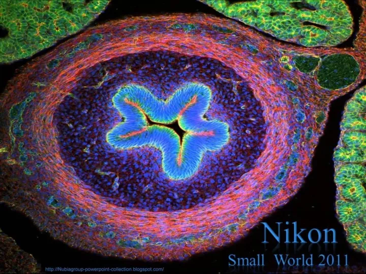
PPS by Nubia_group - https://nubiagroup-powerpoint-collection.blogspot.com/2011/10/nikon-small-world-2011.html

E N D
1st Place, 2011 Dr. Igor Siwanowicz Max Planck Institute of Neurobiology Martinsried, Germany Portrait of a Chrysopa sp. (green lacewing) larva (20x)
2nd Place, 2011 Dr. Donna Stolz University of Pittsburgh Pittsburgh, Pennsylvania, USA Blade of grass (200x)
3rd Place, 2011 Frank Fox Fachhochschule Trier Trier, Rheinland-Pfalz, Germany Melosira moniliformis, living specimen (320x)
4th Place, 2011 Dr. Robin Young The University of British Columbia Vancouver, British Columbia, Canada Intrinsic fluorescence in Lepidozia reptans (liverwort) (20x)
Nikon Small World 5th Place, 2011 Alfred Pasieka Hilden, Germany Microchip surface, 3D reconstruction (500x)
6th Place, 2011 Dennis Callahan California Institute of Technology Department of Applied Physics & Materials Science Pasadena, California, USA Cracked gallium arsenide solar cell films (50x)
Nikon Small World 7th Place, 2011 Gabriel Luna UC Santa Barbara, Neuroscience Research Institute Santa Barbara, California, USA Retinal flatmount of mouse nerve fiber layer (40x)
8th Place, 2011 Dr. Bernardo Cesare Department of Geosciences University of Padua Padova, Italy Graphite-bearing granulite from Kerala (India) (2.5x)
Nikon Small World 9th Place, 2011 Dr. Jan Michels Christian-Albrechts- Universität zu Kiel Kiel, Germany Temora longicornis (marine copepod), ventral view (10x)
10th Place, 2011 Joan Röhl Institute for Biochemistry and Biology University of Potsdam Potsdam, Germany Daphnia magna (freshwater water flea) (100x)
11th Place, 2011 Dr. Jan Michels Christian-Albrechts-Universität zu Kiel Kiel, Germany Ant head, frontal view (10x)
12th Place, 2011 Thomas Deerinck National Center for Microscopy and Imaging Research University of California, San Diego La Jolla, California, USA HeLa (cancer) cells (300x)
13th Place, 2011 Stephen S. Nagy, M.D. Montana Diatoms Helena, Montana, USA Curare vine in cross-section, Chondrodendron tomentosum (45x)
Nikon Small World14th Place, 2011 Yanping Wang Beijing Planetarium Beijing, China Sand (4x)
15th Place, 2011 James H. Nicholson Coral Culture and Collaborative Research Facility, NOAA/NOS/NCCOS/CCEHBR & HML Charleston, South Carolina, USA Porites lobata (lobe coral), live specimen displaying tissue pigmentation response with red fluorescence (12x)
16th Place, 2011 Dr. Christopher Guérin VIB (Flanders Institute of Biotechnology) Ghent, Belgium Cultured cells growing on a bio-polymer scaffold (63x)
17th Place, 2011 Dr. Witold Kilarski EPFL-Laboratory of Lymphatic and Cancer Bioengineering Lausanne, Switzerland Litomosoides sigmodontis (filaria worms) inside lymphatic vessels of the mouse ear (150x)
18th Place, 2011 Benjamin Blonder and David Elliott University of Arizona Department of Ecology & Evolutionary Biology Tucson, Arizona, USA Venation network of young Populus tremuloides (quaking aspen) leaf (4x)
Nikon Small World 19th Place, 2011 Dr. Donna Stolz University of Pittsburgh Pittsburgh, Pennsylvania, USA Mammalian cell collage stained for various proteins and organelles, assembled into a wreath (200-2000x)
20th Place, 2011 Douglas Moore University of Wisconsin - Stevens Point Stevens Point, Wisconsin, USA Agatized dinosaur bone cells, unpolished, ca. 150 million years old (42x)
Honorable Mention, 2011 Jose R. Almodovar Microscopy Center, Biology Department, UPR Mayaguez Campus Mayaguez, Puerto Rico, USA Utricularia gibba (bladderwort) bladder (40x)
Nikon Small World Honorable Mention, 2011 Dr. Paul Appleton University of Dundee Dundee, Scotland, UK Mouse small intestine, stem cells in crypts (40x)Interference Contrast
Nikon Small World Honorable Mention, 2011 Dr. Pedro Barrios-Perez National Research Council of Canada/Institute for Microstructural Sciences Ottawa, Ontario, Canada Gallium antimonide semiconductor wafer surface after metal peel-off (200x)
Nikon Small World Honorable Mention, 2011 Dr. Jorge Bernardino de la Serna MEMPHYS - Center for Biomembrane Physics, Department of Biochemistry and Molecular Biology University of Southern Denmark Odense, Denmark Giant liposomes of pulmonary surfactant (40x)
Nikon Small World Honorable Mention, 2011 Gerd A. Guenther Duesseldorf, Germany Trichodina pediculus (freshwater ciliates), ventral view, living specimens (1000x)
Nikon Small World Honorable Mention, 2011 Gerd A. Guenther Duesseldorf, Germany Nassula ornata (freshwater ciliate), conjugation, living specimens (630x)
Nikon Small World Honorable Mention, 2011 Dr. Marta Guervos Image Processing Unit, Scientific-Technical Facilities, University of Oviedo Asturias, Spain Acacia dealbata (Silver Wattle) pollen grains (40x)
Nikon Small World Honorable Mention, 2011 Charles Krebs Charles Krebs Photography Issaquah, Washington, USA Hydra sp. capturing water flea (40x)
Nikon Small World Honorable Mention, 2011 Dr. David Maitland www.davidmaitland.com Feltwell, UK Enormous dorsal (10x)
Nikon Small World Honorable Mention, 2011 Poulomi Ray Department of Biological Sciences, Clemson University Clemson, South Carolina, USA Chick embryo intestine (20x)
Nikon Small World Honorable Mention, 2011 Kevin Smith MetPrep Ltd Warwickshire, UK Charge coupled device (CCD) sensor, direct surface view (1000x)
Honorable Mention, 2011 Wim van Egmond Micropolitan Museum Rotterdam, Netherlands Argulus (fish louse) (60x)
Nikon Small World Honorable Mention, 2011 Yanping Wang Beijing Planetarium Beijing, China Snowflake (4x)
Honorable Mention, 2011 Dr. Torsten Wittmann University of California, San Francisco California, USA Bovine pulmonary artery endothelial (BPAE) cells fixed and stained for actin, mitochondria, and DNA (60x)
Image of Distinction, 2011 Jose R. Almodovar - Fresh water shrimp eye and head (2x) Microscopy Center, Biology Department, UPR Mayaguez Campus Mayaguez, Puerto Rico, USA
image of Distinction, 2011 - Primary rat neurons grown as neurospheres, observed by Dr. Rowan Orme of Keele University, Keele, UK. (Dr. Rowan Orme)
image of Distinction, 2011 Using laser-triggered high-speed macrophotography, Dr. John H. Brackenbury of the University of Cambridge in Cambridge, UK, captured this image of a water droplet containing a pair of mosquito larvae. (Dr. John H. Brackenbury)
Nikon Small World image of Distinction, 2011 Massimo Brizzi Microcosmo Italia Empoli, Firenze, Italy Ascaris sp. (parasitic nematode), female (150x)
Nikon Small World image of Distinction, 2011 A three dimensional view of a cell culture of breast cancer cells, by Dr. Jonatas Bussador do Amaral and Dr. Gláucia Maria Machado Santelli of the University of São Paulo in São Paulo, Brazil. (Dr. Jonatas Bussador do Amaral, Dr. Gláucia Maria Machado Santelli)
Nikon Small World image of Distinction, 2011 Dr. Douglas Clark of San Francisco, California submitted this image of the dried wing scales of a butterfly (Cethosia biblis) in incandescent light. (Dr. Douglas Clark)
Nikon Small World image of Distinction, 2011 Jonathan Franks of the University of Pittsburgh in Pittsburgh, Pennsylvania, used autofluorescence to observe this algae biofilm. (Jonathan Franks)
Nikon Small World image of Distinction, 2011 John Gaynes University of Utah Salt Lake City, Utah 3 day post-fertilization zebrafish embryo Confocal
Nikon Small World Image of Distinction, 2011 David Millard Austin, Texas, USA Vanessa atalanta (Red admiral butterfly) egg in Urtica dioica (Stinging nettle) trichomes (10x)
Nikon Small World image of Distinction, 2011 The embryonic pectoral fin of Chiloscyllium plagiosum, the Whitespotted bamboo shark, observed by Dr. Andrew Gillis, University of Cambridge, Cambridge, UK. (Dr. Andrew Gillis)
Nikon Small World image of Distinction, 2011 Charles Krebs from Issaquah, Washington brings us this portrait of a water boatman (Corixidae sp.), viewed in reflected light. (Charles Krebs)
Nikon Small World image of Distinction, 2011 Debora Leite of the University of Sao Paulo, in Sao Paulo, Brazil observed this cross-section of the structure of a sugarcane root. (Debora Leite)
Nikon Small World image of Distinction, 2011 Wolfgang Bettighofer Kiel, Germany Closterium lunula (green alga), living specimen from a bog pond Differential Interference Contrast
Nikon Small World image of Distinction, 2011 A naturally formed frost crystal that had grown overnight on a fence in -15 degrees C weather. Image from Jesper Grønne of Silkeborg, Denmark. (Jesper Grønne)