Download
1 / 2
22 likes | 35 Views
OptraSCAN's Solution for Immuno-oncology: Optra PD-L1 automated image analysis for research use supports the interpretation of PD-L1 IHC expression on both tumor and immune cells in solid tumors.<br>Tel : 1-408-524-5300<br>Contact us at- info@optrascan.com<br>Visit- https://www.optrascan.com/solutions/immuno-oncology-pd-l1-biomarker-analysis

E N D
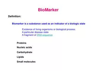
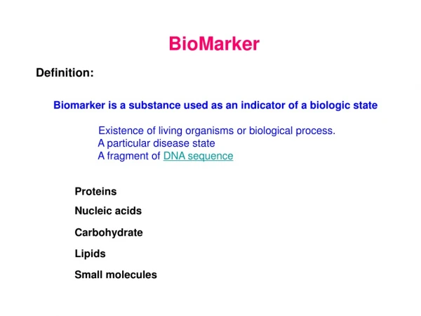
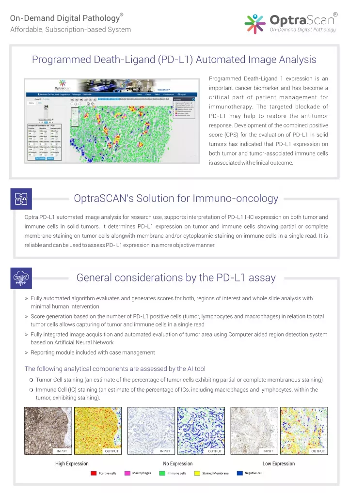
® ® OptraScan On-Demand Digital Pathology Affordable, Subscription-based System Programmed Death-Ligand (PD-L1) Automated Image Analysis Programmed Death-Ligand 1 expression is an important cancer biomarker and has become a critical part of patient management for immunotherapy. The targeted blockade of PD-L1 may help to restore the antitumor response. Development of the combined positive score (CPS) for the evaluation of PD-L1 in solid tumors has indicated that PD-L1 expression on both tumor and tumor-associated immune cells is associated with clinical outcome. OptraSCAN's Solution for Immuno-oncology Optra PD-L1 automated image analysis for research use, supports interpretation of PD-L1 IHC expression on both tumor and immune cells in solid tumors. It determines PD-L1 expression on tumor and immune cells showing partial or complete membrane staining on tumor cells alongwith membrane and/or cytoplasmic staining on immune cells in a single read. It is reliable and can be used to assess PD- L1 expression in a more objective manner. General considerations by the PD-L1 assay Ø Fully automated algorithm evaluates and generates scores for both, regions of interest and whole slide analysis with minimal human intervention Ø Score generation based on the number of PD-L1 positive cells (tumor, lymphocytes and macrophages) in relation to total tumor cells allows capturing of tumor and immune cells in a single read Ø Fully integrated image acquisition and automated evaluation of tumor area using Computer aided region detection system based on Artificial Neural Network Ø Reporting module included with case management The following analytical components are assessed by the AI tool m Tumor Cell staining (an estimate of the percentage of tumor cells exhibiting partial or complete membranous staining) m Immune Cell (IC) staining (an estimate of the percentage of ICs, including macrophages and lymphocytes, within the tumor, exhibiting staining). INPUT INPUT INPUT OUTPUT OUTPUT OUTPUT High Expression No Expression Low Expression Macrophages Nega?ve cell Posi?ve cells Immune cells Stained Membrane
® OptraScan ® On-Demand Digital Pathology Solutions OS-15 OS-FS OS-FL OS-120 15-slide brightfield 7-slide frozen sections, with live view mode 15-slide fluorescence, with 6 filter cubes 120-slide brightfield ® IMAGEPath Web-based Image Management and Viewing TM OptraASSAYS TM TELEPath Web and Mobile Digital Conferencing On-Demand Image Analysis ® CLOUDPath Laboratory Information Management System 100 Century Center Court, Suite 410, San Jose, CA 95112 OptraSCAN is an ISO13485 certified company *All OptraSCAN systems and solutions are for research use only