Download
1 / 4
43 likes | 50 Views
OptraSCANu2019s digital cytology imaging solution will assist pathologists and cytologists to scan, index, and analyze<br>the cytology slides.<br><br>Tel : 1-408-524-5300<br>Contact us at- info@optrascan.com<br>Visit- https://www.optrascan.com/solutions/cytology-imaging

E N D
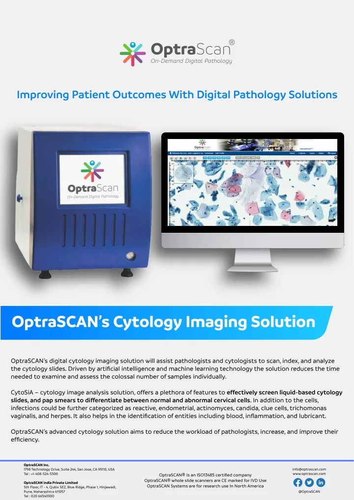
Improving Patient Outcomes With Digital Pathology Solutions OptraSCAN’s Cytology Imaging Solution OptraSCAN’s digital cytology imaging solution will assist pathologists and cytologists to scan, index, and analyze the cytology slides. Driven by artificial intelligence and machine learning technology the solution reduces the time needed to examine and assess the colossal number of samples individually. CytoSiA – cytology image analysis solution, offers a plethora of features to effectively screen liquid-based cytology slides, and pap smears to differentiate between normal and abnormal cervical cells. In addition to the cells, infections could be further categorized as reactive, endometrial, actinomyces, candida, clue cells, trichomonas vaginalis, and herpes. It also helps in the identification of entities including blood, inflammation, and lubricant. OptraSCAN’s advanced cytology solution aims to reduce the workload of pathologists, increase, and improve their efficiency. OptraSCAN Inc. 1798 Technology Drive, Suite 244, San Jose, CA 95110, USA Tel : +1-408-524-5300 info@optrascan.com www.optrascan.com OptraSCAN® Is an ISO13485 certified company OptraSCAN® whole slide scanners are CE marked for IVD Use OptraSCAN Systems are for research use In North America OptraSCAN India Private Limited 5th Floor, IT - 4, Qubix SEZ, Blue Ridge, Phase 1, Hinjewadi, Pune, Maharashtra 411057 Tel : 020 66540900 @OptraSCAN
OptraSCAN’s Z-Stack And Extended Depth Of Field Volume Scan Solution For Cytology Z-Stacking Technology: Stack of images is generated at different focal planes along the z- axis Viewing software enables the user to navigate, zoom up and down the different planes to detect the three-dimensional regions in focus Can scan up-to 7 layers Features Of OptraSCAN’s Volume Scanning Technology: Fully automated multilayer scanning at different focal depths. Automated and User configurable focal intervals for Z stacking Ideal for Rapid On-site evaluation for adequacy of cytology samples (Rapid on-site evaluation (ROSE) Extended Depth Of Field: OptraSCAN’s extended depth of field algorithm generates a single, entirely focused composite image to screen an entire image effectively, easily, and systematically. Advantages: Relatively smaller file size than multiplane images Quicker to acquire Effective and easy navigation for accurate interpretation, sharing and archiving OptraSCAN Inc. 1798 Technology Drive, Suite 244, San Jose, CA 95110, USA Tel : +1-408-524-5300 info@optrascan.com www.optrascan.com OptraSCAN® Is an ISO13485 certified company OptraSCAN® whole slide scanners are CE marked for IVD Use OptraSCAN Systems are for research use In North America OptraSCAN India Private Limited 5th Floor, IT - 4, Qubix SEZ, Blue Ridge, Phase 1, Hinjewadi, Pune, Maharashtra 411057 Tel : 020 66540900 @OptraSCAN
OptraSCAN’s AI & ML- based Image Analysis Solution for Cytology Biomarker Features: Automated computation of sample adequacy for the whole slide cytology image. Identification of abnormal cells and other entities based on morphological features and AI based classification. Identification of reactive, endometrial, actinomyces, candida, clue cells, trichomonas vaginalis, and herpes entities. Identification of entities including blood, inflammation, and lubricant. High Resolution Whole Slide Screening Identification of Infections & Abnormal Cells Faster identification of abnormal cells OptraSCAN Inc. 1798 Technology Drive, Suite 244, San Jose, CA 95110, USA Tel : +1-408-524-5300 info@optrascan.com www.optrascan.com OptraSCAN® Is an ISO13485 certified company OptraSCAN® whole slide scanners are CE marked for IVD Use OptraSCAN Systems are for research use In North America OptraSCAN India Private Limited 5th Floor, IT - 4, Qubix SEZ, Blue Ridge, Phase 1, Hinjewadi, Pune, Maharashtra 411057 Tel : 020 66540900 @OptraSCAN
Identification of Abnormal Cells Identification of Infections (Actinomyces) Identification of HSIL OptraSCAN Inc. 1798 Technology Drive, Suite 244, San Jose, CA 95110, USA Tel : +1-408-524-5300 info@optrascan.com www.optrascan.com OptraSCAN® Is an ISO13485 certified company OptraSCAN® whole slide scanners are CE marked for IVD Use OptraSCAN Systems are for research use In North America OptraSCAN India Private Limited 5th Floor, IT - 4, Qubix SEZ, Blue Ridge, Phase 1, Hinjewadi, Pune, Maharashtra 411057 Tel : 020 66540900 @OptraSCAN