Download
1 / 3
32 likes | 39 Views
OptraSCAN Fluorescence Scanning & Analysis is a Small Footprint, Automated Whole Slide Fluorescence & Brightfield Scanning With High-Resolution Imaging.<br>Visit-https://optrascan.com/scan/os-fl-multiplexing-fluorescence-scanner/ <br>Contact us at-info@optrascan.com

E N D
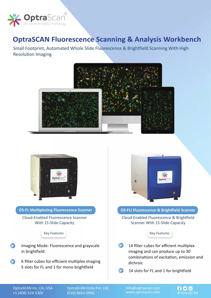
OptraSCAN Fluorescence Scanning & Analysis Workbench Small Footprint, Automated Whole Slide Fluorescence & Brigh�ield Scanning With High Resolu�on Imaging OS-FL Mul�plexing Fluorescence Scanner OS-FLi Fluorescence & Brigh�ield Scanner Cloud-Enabled Fluorescence Scanner With 15-Slide Capacity Cloud-Enabled Fluorescence & Brigh�ield Scanner With 15-Slide Capacity Key Features Key Features Imaging Mode: Fluorescence and grayscale in brigh�ield 14 filter cubes for efficient mul�plex imaging and can produce up to 30 combina�ons of excita�on, emission and dichroic 6 filter cubes for efficient mul�plex imaging 5 slots for FL and 1 for mono brigh�ield 14 slots for FL and 1 for brigh�ield OptraSCAN Inc. CA, USA +1 (408) 524-5300 OptraSCAN India Pvt. Ltd. (020) 6654-0900 info@optrascan.com www.optrascan.com @OptraSCAN
An ISO 13485 cer�fied company OptraSCAN systems are CE mark for IVD use All OptraSCAN systems are for research use only in North America Technical Specifica�ons User friendly, intui�ve LED touchscreen Compa�ble with mul�ple image formats: JPEG2000, TIFF, SVS, MRXS, CZI, NDPI, JPEG, OpenSlide compa�ble Magnifica�on: 20x or 40x magnifica�on Resolu�on: 0.50 µm/pixel at 20x, 0.25 µm/pixel at 40x 15 slide capacity Dimensions & Weight of OS-FL - Approximate Width- 16 ½ ʺ, Length- 14ʺ, Height- 16ʺ, Weight- 60lbs Slide Formats: Standard 25x75mm (1ʺx3ʺ) slides Bar code and case reconcilia�on Image capture region: 25x50mm Dimensions & Weight of OS-FLi - Approximate Width- 14 ʺ, Length- 24ʺ, Height- 19ʺ, Weight- 88lbs Image Storage Space: approx. 300 MB for a single channel for a 15mm x 15 mm �ssue Opera�ng System: Windows 7, 8, 8.1, 10 IMAGEPath: Image Management System included for viewing, storing and archiving Data Storage 1-10 TB TELEPath: Telepathology included for real-�me, remote consulta�ons FL Viewer IHC Mul�plex So�ware Key Features Large image support Illumina�on correc�on, vigne�ng correc�on Photobleaching correc�on Pixel to pixel spa�al registra�on Individual signal op�miza�on Spectral unmixing Mul�-level cell segmenta�on Ga�ng to construct cells from segmented cellular parts Robust quan�ta�ve analysis for each imaged channel Comprehensive feature extrac�on 3D reconstruc�on Image manipula�ons Brightness | Contrast and opacity Custom channel naming Layer blending Image opera�ons Atlas mapping Pan-and-zoom func�onality for high resolu�on images Drawing & impor�ng of user-defined regions of interest Technical Specifica�ons Supports CZI, BigTIFF, JP-2000 and standard TIFF with no restric�on on image size & number of channels Features associated with each cellular object is computed & available for viewing and analysis So�ware is na�vely compa�ble and seamlessly integrated to support end-to-end image data processing and analysis mul�plexed fluorescent images Segmented cells are displayed in a cell tray Opera�ng system: Windows 7, 8, 8.1, 10 OptraSCAN Inc. CA, USA +1 (408) 524-5300 OptraSCAN India Pvt. Ltd. (020) 6654-0900 info@optrascan.com www.optrascan.com @OptraSCAN
An ISO 13485 cer�fied company OptraSCAN systems are CE mark for IVD use All OptraSCAN systems are for research use only in North America So�ware provides precise pixel-to-pixel spa�al registra�on for all imaged channels per specimen, including those sequen�ally acquired a�er repeated an�body tripping, restaining and reimaging Mul�-Level Cell Segmenta�on Detec�on algorithms to iden�fy and classify cellular en��es Algorithms can be fine tuned by user 3D Re-Construc�on Selec�on of mul�ple sec�ons Fetching of composite segmented cell and process objects that need to be reconstructed 3 Dimensional (3-D) visualiza�on Ga�ng Module Morphological opera�ons between segmented objects in different channels to reconstruct cells Addi�on and subtrac�on of segmented objects between two or more channels supported Data Export In FCS & ICE So�ware supports FCS and ICE export file formats compa�ble with 3rd party flow cytometry and image cytometry so�wares Pan-And-Zoom Func�onality For High Resolu�on Images Real-�me pan and zoom So�ware supports func�onality of drawing user adjustable ROI’s: dropdown/ select the ROI op�on for selec�ng a par�cular area in the input image The so�ware supports adding annota�ons for the color channels (square, rectangle, circle, ellipsoid, polygonal, freeform) to classify and compare the data across mul�ple areas of interest The so�ware supports func�onality to save the ROI’s drawn on the image OptraSCAN Inc. CA, USA +1 (408) 524-5300 OptraSCAN India Pvt. Ltd. (020) 6654-0900 info@optrascan.com www.optrascan.com @OptraSCAN
