Download
1 / 7
70 likes | 76 Views
The market is flooded with different kinds of medical imaging software for viewing DICOM images. This includes free medical imaging software as well as premium software that may offer more advanced features. As radiologists become accustomed to the latest medical imaging software for viewing and storing images, manufacturers are turning their attention to other areas of the imaging workflow, identifying problems that need to be solved and seeing if they can come up with innovative solutions for the same. In this article, we review the different types of medical imaging software that have been designed to do more than just view DICOM medical images.

E N D
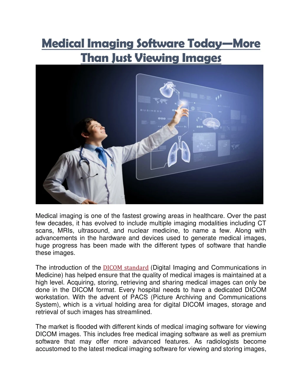
Medical Imaging Software Today—More Than Just Viewing Images Medical imaging is one of the fastest growing areas in healthcare. Over the past few decades, it has evolved to include multiple imaging modalities including CT scans, MRIs, ultrasound, and nuclear medicine, to name a few. Along with advancements in the hardware and devices used to generate medical images, huge progress has been made with the different types of software that handle these images. The introduction of the DICOM standard (Digital Imaging and Communications in Medicine) has helped ensure that the quality of medical images is maintained at a high level. Acquiring, storing, retrieving and sharing medical images can only be done in the DICOM format. Every hospital needs to have a dedicated DICOM workstation. With the advent of PACS (Picture Archiving and Communications System), which is a virtual holding area for digital DICOM images, storage and retrieval of such images has streamlined. The market is flooded with different kinds of medical imaging software for viewing DICOM images. This includes free medical imaging software as well as premium software that may offer more advanced features. As radiologists become accustomed to the latest medical imaging software for viewing and storing images,
manufacturers are turning their attention to other areas of the imaging workflow, identifying problems that need to be solved and seeing if they can come up with innovative solutions for the same. In this article, we review the different types of medical imaging software that have been designed to do more than just view DICOM medical images. Medical Image Analysis Software Any software that can ‘analyze’ data that is obtained from medical images is referred to as medical image analysis software. Analysis can take the form of aiding diagnosis, comparing images between patients or within the same patient at different time points to assess the progress of disease, and evaluating prognosis. In conjunction with the improvement in imaging technology, great strides are being made with regards to the analytic ability of medical imaging software, in the effort to create software capable of independently detecting clinical anomalies in medical images. What is the need for medical image analysis software? Analysis is usually a cognitive function that is performed by the radiologist or physician who views the medical image. With advances in healthcare, the number of scans being requested for patients has skyrocketed. Medical scan outputs today are available in greater detail and in multiple sections, leading to a greater number of images that need examination. Interpretation of so many images by a radiologist not only requires tremendous skill, it is also time-consuming and exhausting. While the workload for radiologists has multiplied over the years, the growth in the number of trained radiologists has only mirrored half the increase in workload. The result is an acute human resource shortage within the context of radiology workload. A proposed solution to this problem is the use of machines to interpret medical images and detect anomalies. Medical image analysis software uses deep learning algorithms to read and evaluate images. It is capable of sifting through hundreds of images at a time, and therefore can handle large workloads. It can be trained to ‘flag’ images with suspicious findings, which can expedite processes for radiologists in the sense that they need not go through all the images and just focus on those that are flagged. What are some medical image analysis software products on the market? •Aidoc: Aidoc, a Tel Aviv based company, has developed medical image analysis software that provides diagnostic support for full body CT scans.
The application analyzes CT scans of the head, neck, chest and abdomen, and is capable of detecting high-level visual abnormalities. One case study conducted by the company demonstrated that using Aidoc significantly reduced report turnaround time, particularly for scans of the head and neck. •Arterys: Arterys is a San Francisco based company that combines deep learning AI algorithms with cloud computing. The medical image analysis software has been shown to increase the speed and accuracy of analysis. Initially developed for cardiac MRIs, Arterys has now developed similar applications for liver MRIs, lung MRIs and mammograms, and helps identify pathological lesions in these regions. What are the limitations of medical image analysis software? At the end of the day, medical image analysis software is as good as the computer algorithms it is built on. A computer does not ‘see’ things and cannot think, and its output is based on a series of numbers and algorithms. The results generated are therefore based on the algorithms that it has been programed with. There is therefore plenty of room for error here as the technology is still nascent. While medical imaging analysis software can certainly reduce radiologist workload, it is not ready yet to completely replace the radiologist. It is still in its infancy and is not used as commonly as its less automated counterpart, the medical image processing software. Medical Image Processing Software Medical image processing software in essence transforms images after they have been acquired. While some groups consider medical image processing software to be a part of medical image analysis software, it does not do much to analyze images. Nevertheless, processing makes the job of manual analysis easier for the radiologist. Medical image processing is of three types—image segmentation, image registration, and image visualization. Image Segmentation Segmentation refers to the process of dividing a single image into small parts or segments. Ideally, these segments need to be meaningful, that is, each segment should depict a different structure or organ.
Medical image segmentation software is capable of performing the following functions: •Locating the region of interest: The software can identify abnormalities in the region of interest, including tumors, nodules and other pathologies. •Discerning anatomical boundaries: Segmentation software can identify the boundaries of body structures such as blood vessels. •Measuring volumes: Medical image segmentation software can be used to calculate the volumes of specific structures such as anatomical cavities or tumors. It is particularly useful to monitor changes in tumor size during the course of treatment. Image Registration Image registration is a process that allows images to be aligned in the correct manner. In this technique, the computer is acquainted with a series of ‘target’ images. When it is fed a new image, this new ‘source’ image is transformed to become similar in alignment to the to the target image. Image registration can be achieved using three methods—transformation models, similarity functions, and optimization procedures. Applications of image registration through medical image processing software: •Image fusion: In image fusion, medical image data that come from different sources can be fused together into a single dataset. This is extremely useful in understanding how anatomy correlates with functional processes. For instance, CT scans provide structural information, while PET scans provide metabolic information. Using image fusion, both sets of information can be obtained through a single dataset. •Studying changes over time: Image registration can be used to compare a series of images over time. This is useful to assess changes within the same imaging session, such as heart movements or respiratory function. It can also be applied to long-term changes, such as monitoring the progression of a disease over a few years. •Characterizing anatomical features: Image registration can also compare images between different subjects in a population. This can be used to characterize anatomical features in a given population.
•Interventional procedures: Computer-assisted surgery is made possible with image registration. By applying the pre-operative CT scan or MRI image to the intraoperative setting, image-guided surgery becomes possible. Image Visualization Medical image visualization software changes the way that the original dataset can be viewed. This allows for analysis from different points of view. Visualization is essentially the process of exploring data, transforming it if needed, and then viewing it with greater depth and clarity in comparison with the original dataset. There are several post-processing techniques that allow for medical image visualization. Applications of image visualization through medical image processing software: •3D reconstruction: 3D medical imaging software is almost always built into regular medical image processing software programs. 3D reconstruction involves addition of all the sections acquired in a single dataset and combining them into a single image. This allows operators to easily interpret abnormalities as there is better anatomical orientation compared to individual sections. 3D medical imaging software also helps in quicker identification of abnormalities. Greater detail can then be visualized with 2D visualization if required. •2D visualization: This is an inverse of the 3D reconstruction technique. It can be used to either display the original imaging data from 3D or 4D reconstructions, or it can be used to view different sections from the original dataset. An example of 2D visualization is multiplanar reformatting, which allows new sections to be made from 3D and 4D reconstructions, at planes that are different from the original planes. MPR finds application in the visualization of curvilinear structures, including the spinal canal and blood vessels. Most kinds of 3D medical imaging software allow for MPR as well. Medical Image Management Software The simultaneous increase in the number of patients undergoing diagnostic medical imaging and the quality of medical images being acquired, which means enormous data files, has led to massive volumes of datasets being handled by
healthcare centers and hospitals. The storage, retrieval and handling of this huge volume of imaging data can be a challenge in itself. Medical image management software makes this process easier by organizing and integrating such datasets. Medical image management software consists of a PACS server that can be integrated with a regular DICOM workstation. A standard medical image management software should have the following features: •Replaces physical archiving by storing all medical image datasets digitally in an organized manner. •Allows radiologists access to medical imaging data from any geographical location, and allows multiple users to view data at the same time on different systems. •Allows exporting of images to other file formats, so that they may be used for teaching, learning, or for disseminating images through publications and websites. •Allows integration of medical image data with patient data in other records, such as the electronic health record, health information system, and radiology information system (RIS). Medical Image Dose Tracking Software A major downside to medical imaging is radiation exposure. Measuring the dose of radiation involved during scan acquisition is now possible with tracking software. Why is medical image dose tracking software necessary? With the increasing use of CT-guided diagnosis and intervention, including nuclear medicine based scans and angiography, there has been a steady rise in both patient and physician exposure to radiation. Statutory bodies have noted this and made it mandatory to track the amount of radiation that patients receive and enter this in their health records. It is also required to track the amount of radiation that doctors are exposed to during the course of their work. What are some available dose tracking applications?
To aid with dose tracking, several developers of medical image management software have come up with solutions. For instance, GE offers a program called DoseWatch. It tracks the radiation dose administered to patients at a given institution. The data can be classified according to the individual device, the protocol, or the operator so that it becomes easy to identify dose outliers. Other applications like Sectra offer web-based dose tracking. Sectra is certified by the American College of Radiologists and can submit dose data from a hospital directly to their Dose Index Registry. PostDICOM—Comprehensive Medical Imaging Solution PostDICOM integrates the medical imaging software functions that we have described above into one feature-packed program. It is sophisticated medical image management software that allows cloud-based storage and retrieval of medical images. PostDICOM is compatible with multiple operating systems, including Windows, Linux, Mac OS and Android. This free medical imaging software offers advanced visualization options and is integrated with medical image segmentation software. The free version allows 50 GB of data storage in the cloud. Additional storage can be purchased at a nominal cost. Visit postdicom.com to learn more about this handy piece of software.