Download
1 / 26
260 likes | 518 Views
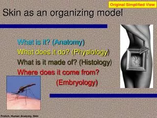
Original Simplified View. Skin as an organizing model. What is it? (Anatomy) What does it do? (Physiology ) What is it made of? (Histology ) Where does it come from? (Embryology). Skin as an organizing model. What is it? (Anatomy) What does it do? (Physiology )

E N D
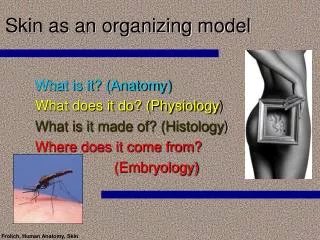
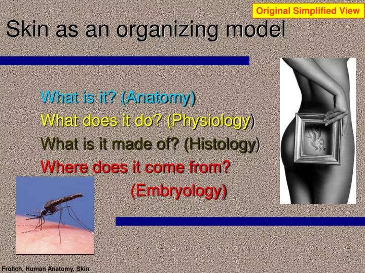
Original Simplified View Skin as an organizing model What is it? (Anatomy) What does it do? (Physiology) What is it made of? (Histology) Where does it come from? (Embryology) Frolich, Human Anatomy, Skin
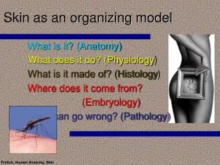
Skin as an organizing model What is it? (Anatomy) What does it do? (Physiology) What is it made of? (Histology) Where does it come from? (Embryology) [brainstorm—what questions remain?] Frolich, Human Anatomy, Skin
Skin revisited: Filling in the Blanks Anatomy—dermatomes and spinal nerves Histology epidermal cells and layers dermal layers other structures Physiology details of skin protection function Pathology of skin skin cancers burns Frolich, Human Anatomy, Skin
Original Simplified View What is it (anatomy)? • Outer covering of body • Keratinized • Hair—head, pubic regions • Nails—fingers, toes • Divided into segments called dermatomes (“Zebra-man”) • CE (clinical example): Shingles Frolich, Human Anatomy, Skin
Dermatomes—more details Dermatomes and spinal nerves Frolich, Human Anatomy, Skin
Dermatomes—more details Dermatomes and segmentation in basic body plan Frolich, Human Anatomy, Skin
Epidermis anatomy—more details Hair and nails—modified structures of epidermis • Nails • Scale-like epidermal structure • Cells bind together and have “hard” keratin • Grows out from root of nail • Hair • Each shaft has three layers of keratinzed cells filled with “hard” keratin • Flat, ribbon-like shaft produces kinky hair; oval shaft makes wavy hair; round shaft makes coarse hair • Hair color due to amount of melanins of different colors made my melanocytes at base of hair follicle; red hair also has iron-containing pigment; gray/white hair due to decreased melanin production • Hair follicle • Fold of epidermal surface into dermis • Hair grows from here • Has nerve plexus to give touch/tickle sensation • Connective tissue sheath derived from dermis • Hair length due to relationship between active and inactive phases of follicle (e.g., eyebrow follicles active only three to four months; head follicles can be continuosly active for years) Frolich, Human Anatomy, Skin
Epidermis anatomy—more details Nails—scale-like epidermis Frolich, Human Anatomy, Skin
Epidermis anatomy—more details Epidermis—more detail Hairs and hair follicles Frolich, Human Anatomy, Skin
Original Simplified View What is it made of?(histology—study of tissues) • Layers of skin are two fundamental types of tissue organization • Epidermis = epithelium • Dermis = connective tissue Frolich, Human Anatomy, Skin
Epidermis histology—more details Layers of epidermis • Stratum basale/germinativum (“basal or “forming” layer) • One layer thick mitotic cells • 10-25% melanocytes with processes into next layer • Merkel cells with sensory neurons • Stratum spinosum (“prickly” layer) • Cells appear spiny due to numerous desmosomes • Many Langerhans cells • Stratum granulosum (“grainy” layer • Cells flatten • Organelles/nuclei begin to disintegrate • Keratin precursor granules begin to form • Lamellated granules with water-proof lipid form and will be spewed out between cells • Stratum corneum (“horny” layer) • Cells are dead—too far from underlying capillaries to live • 20-30 cells thick up to ¾ of dermal thickness • Keratin, thickened membranes and glycolipids between cells provide “overcoat” for body to protect against water loss and other possible “assaults” on body Frolich, Human Anatomy, Skin
Epidermis histology—more details Cells in epidermis • Keratinocytes—epidermal cells that make keratin • Merkel cells—associated with touch sensory neurons • Langerhans cells—macrophages (from dermis) migrate in to form spider-like immune barrier • Melanocytes (at border with dermis) make pigment to give skin color Frolich, Human Anatomy, Skin
Epidermis histology—more details Epidermis—a stratified epithelium Frolich, Human Anatomy, Skin
Original Simplified View Dermis—simple view Frolich, Human Anatomy, Skin
Dermis histology—more details Dermal layers • Papillary layer (layer with “nipples” or tiny projections) • Thin superficial layer of areolar connective tissue • Papillae house capillary loops, nerve endings, sweat glands • In hands and feet papillae on dermal ridges forming fingerprint patterns • Reticular layer • 80% of thickness • Dense irregular collagen fibers, mostly parallel to skin surface • Predominant direction of fibers forms cleavage or tension lines in skin (important for incisions) • Flexure lines in skin at joints where reticular layer is bound to underlying connective tissues to provide “give” when joint flexes Frolich, Human Anatomy, Skin
Epidermis/Dermis histology—more details Glands and keratinized appendages: Is it dermis…or epidermis? • Sebaceous glands • Clumps of epithelial tissue distributed within dermis • Secrete “sebum”—oily, fat-based substance that is also anti-bacterial • Located all over body • Sweat glands • Microscopic clumps of epithelial tissue distributed within dermis, duct extends out through dermis to pore (not “pores” of face complexion which are hair follicles) • More than 2.5 million glands per person • Eccrine sweat glands, concentrated on hands and soles of feet and forehead, secrete sweat to cool body, also “cold sweat” of fear, emotion. • Apocrine glands, concentrated in armpits and groin, analogous with sexual scent glands of other animals, odor comes from bacteria that concentrate here. • Ceruminous glands: modified sweat glands in ear canal produce ear wax • Mammary glands: modified sweat glands in female breast produce mother’s milk Frolich, Human Anatomy, Skin
Epidermis/Dermis histology—more details Skin Glands Frolich, Human Anatomy, Skin
Original Simplified View What does it do? • PROTECTION • Air = desiccation • Water = bloating/”pruning” • Sun/u-v = burns • Cold/heat = frostbite/heat stroke/thermoregulation • STRUCTURAL • Integrity of body • Muscle attachment Frolich, Human Anatomy, Skin
Physiology—more details What does it do? • PROTECTION • Chemical barriers—”acid mantle” from low “pH of skin secretions retards bacterial growth • Water-proofing from lipid secretions of epidermis • Melanin from melanocytes and DNA in all skin cells prevents burning from sun • Immune cells in dermis and epidermis fight infection and present antigens to other immune cells • Evaporation of sweat cools body • Closing of dermal capillaries prevents heat loss • Supplement liver in “disarming” cancer-causing chemicals that penetrate epidermis • SENSATION—nerve endings • METABOLIC FUNCTION—vitamin D synthesis Frolich, Human Anatomy, Skin
Physiology—more details Pathology of skin • Skin cancer (what are the cells involved?) • Basal cell carcinoma (a) • Common and benign, 30% of white people get, slow-growing and easily incised surgically • Squamous cell carcinoma (b) • Less common, faster-growing but still easily removed surgically • Melanoma (c) • Highly metastatic (spreads rapidly) • Related to U-V exposure • Early detection key—can start in retina (importance of ophthalmologic exams) Frolich, Human Anatomy, Skin
Physiology—more details Frolich, Human Anatomy, Skin
Physiology—more details Pathology of skin Burns • Rule of nines • Degree of burns • Synthetic skin from silicon epidermis and spongy dermis made of collagen fibers and ground cartilage Frolich, Human Anatomy, Skin
Physiology—more details Interrelationship with other systems • Systems are not isolated, but just one way of analyzing how body works • Skin is tightly linked to other systems of body • Skeletal: Skin forms “exoskeleton”—stiff attachment point for muscles; also synthesizes Vitamin D necessary for calcium deposit process • Muscular: Many muscles attach on the skin; muscles lose heat through skin • Nervous: Sensory cells (Merkel cells) transmit touch sensation to neurons; blood vessel, sweat, arrector pili muscles controlled by nervous system • Cardiovascular: Skin as blood reservoir, heat exchange organ • Immune: Very important role of skin as “first barrier” with Langerhans cells and macrophages as cellular immune response; edema or swelling common in infected skin regions • Reproduction: skin responds to erotic stimuli; mammary glands are highly modified sweat glands in skin; stretching of skin to accomondate fetus during pregnancy Frolich, Human Anatomy, Skin
New Topic of Interest Electronic Skin Frolich, Human Anatomy, Skin
New Topic of Interest Distribution of skin Color • Skin color around world Frolich, Human Anatomy, Skin
New Topic of Interest Genetics of skin color From Genetics Chapter On-Line Biology Book Frolich, Human Anatomy, Skin