Download
1 / 44
460 likes | 1.15k Views
FLUID AND ELECTROLYTE BALANCES Ms. Ida Anitha Lecturer College of Nursing CMC, Vellore. WHY IS IT IMPORTANT FOR NURSES TO KNOW ABOUT FLUID & ELECTROLYTE BALANCE. INTRODUCTION. Water is found everywhere on earth including human body In an adult 60% of the weight is water

E N D
FLUID AND ELECTROLYTE BALANCESMs. Ida AnithaLecturerCollege of NursingCMC, Vellore
WHY IS IT IMPORTANT FOR NURSES TO KNOW ABOUT FLUID & ELECTROLYTE BALANCE
INTRODUCTION • Water is found everywhere on earth including human body • In an adult 60% of the weight is water • Two third of the body’s water is found in the cell
DISTRIBUTION OF BODY FLUIDS Body fluids are distributed in two distinct compartments: 1.Extracellular fluids[ECF] Which includes interstitial fliud & intravascular fluid 2.Intracellular fluids[ICF]
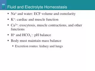
COMPOSITION OF BODY FLUIDS The fluids circulating throughout the body in extracellular and intracellular fluid spaces contain 1.Electrolytes 2.Minerals 3.Cells
MOVEMENT OF BODY FLUIDS • Diffusion • Osmosis • Filtration • Active transport
REGULATION OF BODY FLUIDS • Fluid intake • Fluid output • Hormonal influence • Lymphatic influences • Neurologic influences • Renal influences
ACID-BASE BALANCE • Chemical regulation • Biologic regulation • Physiological regulation 1.Lungs 2.Kidneys
FLIUD IMBALANCES The five types of fluid imbalances that may occur are: • Extracellular fluid imbalances(EVFVD) • Extracellular fluid volume excess(ECFVE) • Extracellular fluid volume shift • Intracellular fluid vloume excess(ICFVE) • Intrcellular fluid volume deficit(ICFVD)
EXTRACELULLAR FLUID VOLUME DEFICIT • An ECFVD, commonly called as dehydration , is a decrease in intravascular and interstitial fluids • An ECFVD can result in cellular fluid loss if it is sudden or severe
THREE TYPES OF ECFVD • Hyperosmolar fluid volume deficit- water loss is greater than the electrolyte loss • Isosmolar fluid volume deficit – equal proportion of fluid and electrolyte loss • Hypotonic fluid volume deficit – electrolyte loss is greater than fluid loss
Severe vomiting Diaphoresis Traumatic injuries Third space fluid shifts [percardial, pleural, pertonial and joint cavities] Fever Gatrointestinal suction Ileostomy Fistulas Burns Hyperventilation Decresed ADH secretions Diabetes insipidus Addison’s disease or adrenal crisis Diuretic phase of acute renal failure Use of diuretics ETIOLOGY AND RISK FACTORS
ELDERLY ARE HIGH RISK OF ECFVD DUE TO • Decreased thirst response • Decreased renal concentration of urine • Altered ADH response • Increased drug – drug interaction • Multiple chronic diseases • Decreased access to fluids due to financial or transportation barriers • Debilitation • Chemical or physical restraint • Changes in mental status
CLINICAL MANIFESTATION • In Mild ECFVD, 1to 2 L of water or 2% of the body weight is lost • In Moderate ECFVD, 3 to 5L of water loss or 5%weight loss • IN Severe ECFVD , 5 to 10 L of water loss or 8% of weight loss
Thirst Muscle weakness Dry mucus membrane;dry cracked lips or furrowed tongue Eyeballs soft and sunken (severe deficit) Apprehension , restlessness, headache , confusion, coma in severe deficit Elevated temperature Tachycardia, weak thready pulse Peripheral vein filling> 5 seconds Postural systolic BP falls >25mm Hg and diastolic fall > 20 mm Hg , with pulse increases > 30 Narrowed pulse pressure, decreased CVP&PCWP Flattened neck veins in supine position Weight loss Oliguria(< 30 mlper hour) Decreased number and moisture in stools CLINICAL MANIFESTATION
LABORATORY FINDINGS • Increased osmolality(> 295 mOsm/ kg) • Increased or normal serum sodium level (> 145mEq/ L ) • Increase BUN (>25 mg / L ) • Hyperglycemia ( >120 mg /dl ) • Elevated hematocrit (> 55%) • Increased specific gravity ( > 1.030)
MANAGEMENT Mild fluid volume loss can be corrected with oral fluid replacement -if client tolerates solid foods advice to take 1200 ml to 1500ml of oral fluids -if client takes only fluids, increase the total intake to 2500 ml in 24 hours
Management of Hyperosmolar fluid volume deficit • Administration of hypotonic IV solution , such as 5% dextrose in 0.2 %saline • If the deficit has existed for more than 24 hours,avoid rapid correction of fluid [sodium solution to be infused at the rate of 0.5 to 0.1m Eq/ L/ hr]
If heamorrhage is the cause for ECFVD • Packed red cells followed by hypotonic IV fluids is administered • In situations where the blood loss is less than 1 L normal saline or ringer lactate may be used • clients with severe ECFVD accompanied by severe heart , liver, or kidney disease cannot tolerate large volumes of fluid and sodium
EXTRACELLULAR FLUID VOLUME EXCESS • ECFVE is increased fluid retention in the intravasular and interstitial spaces
ETIOLOGY AND RISK FACTORS • Heart failure • Renal disorders • Cirrhosis of liver • Increased ingestion of high sodium foods • Excessive amount of IV fluids containing sodium • Electrolyte free IV fluids • SIADH,Sepsis • decreased colloid osmotic pressure • lymphatic and venous obstruction • Cushing’s syndrome & glucocorticoids
CLINICAL MANIFESTATION • Constant irritating cough • Dyspnea & crackles in lungs • Cyanosis, pleural fffusion • Neck vein obstruction • Bounding pulse &elevated BP • S3 gallop • Pitting & sacral edema • Weight gain • Increased CVP& PCWP • Change in level of consiousness
LAB INVESTIGATION • serum osmolality <275mOsm/ kg • Low , normal or high sodium • Decreased hematocrit [ < 45%] • Specific gravity below 1.010 • Decreased BUN [< 8mg/ dl]
MANAGEMENT • Diuretics [combination of potassium sparing and potassium depleting diuretics] • In people with CHF, ACE inhibitors and low dose of beta blockers are used • A low sodium diet
EXTRACELLULAR FLUID VOLUME SHIFT: THIRD SPACING • Fluid that shifts into the interstitial spaces and remain there is called as third space fluid • Common sites are abdomen , pleural cavity, peritoneal cavity and pericardial sac
RISK FACTORS • Crushing injuries, major tissue trauma • Major surgery • Extensive burns • Acid –base imbalances and sepsis • Perforated peptic ulcers • Intestinal obstruction • Lymphatic obstruction • Autoimmune disorders • Hypoalbunemia • GI tract malabsorption
CLINICAL MANIFESTATION • skin pallor • Cold extremities • Weak and rapid pulse • Hypotension • Oliguria • Decreased levels of consiousness LAB INVESTIGATION • Elevated hematocrit & BUN level
MANAGEMENT Treat the cause • For burns and tissue injuries large volume of isosmolar IV fluid is administered • Albumin is administered for protein deficit • IV fluid intake is maintained after major surgery to maintain kidney perfusion • Pericardiocentesis if pericarditis is the result • Paracentesis for ascitis
INTRACELLULAR FLUID VOULME EXCESS:WATER INTOXICATION • ICFVE is increase in amount of water inside the cells
ETIOLOGY • Administration of excessive amount of hyposmolar IV fluids[0.45%saline or 5%dextrose in water] • Consumption of excessive amount of tap water without adequate nutritional intake • SIADH • Schizophrenia[compulsive water consumption]
CLINICAL MANIFESTATIONS • Headaches • Behavioral changes • Apprehension • Irritability, disorientation and confusion • Increased ICP – pupillary changes and decreased motor and sensory function • Bradycardia, elevated BP, widened pulse pressure & altered respiratory patterns, Babinski’s response flaccidity, projectile vomiting, Papilledema, delirium, convulsions &coma
LABORATORY FINDINGS • High serum sodium level- 125 mEq/L • decreased hamatocrit
MANAGEMENT • Early administration of IV fluids containing sodium chloride cam prevent SIADH • oral fluids such as juices or soft drinks can be given orally every hour • Perform neurologic checks every hour to see if cranial changes are present • Monitor fluid intake , IV fluids and fluid output hourly and weight daily • Administer antiemetics for food and fluid retention
INTRACELLULAR FLUID VOLUME DEFICIT • Severe hypernatremia and dehydration can cause ICFVD • Relatively rare in healthy adults • common in elderly people and in those conditions that result in acute water loss • Symptoms include confusion, coma, and cerebral hemorrhage
Sodium imbalances Definition Risk factors/ etiology Clinical manifestation Laboratory findings management Hyponatr -aemia It is defined as a plasma sodium level below 135 mEq/ L Kidney diseases Adrenal insufficiency Gastrointestinal losses Use of diuretics (especially with along with low sodium diet) Metabolic acidosis • Weak rapid pulse • Hypotension • Dizziness • Apprehension and anxiety • Abdominal cramps • Nausea and vomiting • Diarrhea • Coma and convulsion • Cold clammy skin • Finger print impression on the sternum after palpation • Personality change • Serum sodium less than 135mEq/ L • serum osmolality less than 280mOsm/kg • urine specific gravity less than 1.010 • Identify the cause and treat • *Administration of sodium orally, by NG tube or parenterally • *For patients who are able to eat & drink, sodium is easily accomplished through normal diet • *For those unable to eat,Ringer’s lactate solution or isotonic saline [0.9%Nacl]is given • *For very low sodium 0.3%Nacl may be indicated • *water restriction in case of hypervolaemia
Sodium imbalan -ce Definition causes Clinical manifestation Lab findings management Hypernat -remia It is defined as plasma sodium level greater than 145mE q/L *Ingestion of large amount of concentrated salts *Iatrogenic administration of hypertonic saline IV *Excess alderosterone secretion · Low grade fever · Postural hypertension · Dry tongue & mucous membrane · Agitation · Convulsions · Restlessness · Excitability · Oliguria or anuria · Thirst · Dry &flushed skin *high serum sodium 135mEq/L *high serum osmolality295mO sm/kg *high urine specificity 1.030 *Administration of hypotonic sodium solution [0.3 or 0.45%] *Rapid lowering of sodium can cause cerebral edema *Slow administration of IV fluids with the goal of reducing sodium not more than 2 mEq/L for the first 48 hrs decreases this risk *Diuretics are given in case of sodium excess *In case of Diabetes insipidus desmopressin acetate nasal spray is used *Dietary restriction of sodium in high risk clients
Potassium imbalances Definition Causes Clinical manifestation Lab findings Management Hypokalemia It is defined as plasma potassium level of less than 3.0 mEq/L *Use of potassium wasting diuretic *diarrhea, vomiting or other GI losses *Alkalosis *Cushing’s syndrome *Polyuria *Extreme sweating *excessive use of potassium free Ivs *weak irregular pulse *shallow respiration *hypotesion *weakness, decreased bowel sounds, heart blocks , paresthesia, fatigue, decreased muscle tone intestinal obstruction * K – less than 3mEq/L results in ST depression , flat T wave, taller U wave * K – less than 2mEq/L cause widened QRS, depressed ST, inverted T wave Mild hypokalemia[3.3to 3.5] can be managed by oral potassium replacement Moderate hypokalemia *K-3.0to 3.4mEq/L need 100to 200mEq/L of IV potassium for the level to rise to 1mEq/ Severe hypokalemia K- less than 3.0mEq/L need 200to 400 mEq/L for the level to rise to l mEq/L *Dietary replacement of potassium helps in correcting the problem[1875 to 5625 mg/day]
Definition Causes Clinical manifestation Lab findings Management Hyperkalemia It is defined as the elevation of potassium level above 5.0mEq/L Renal failure , Hypertonic dehydration, Burns& trauma Large amount of IV administration of potassium, Adrenal insufficiency Use of potassium retaining diuretics & rapid infusion of stored blood Irregular slow pulse, hypotension, anxiety, irritability, paresthesia, weakness *High serum potassium 5.3mEq/L results in peaked T wave HR 60 to 110 *serum potassium of 7mEq/L results in low broad P- wave *serum potassium levels of 8mEq/L results in no arterial activity[no p-wave] *Dietary restriction of potassium for potassium less than 5.5 mEq/L *Mild hyperkalemia can be corrected by improving output by forcing fluids, giving IV saline or potassium wasting diuretics *Severe hyperkalemia is managed by 1.infusion of calcium gluconate to decrease the antagonistic effect of potassium excess on myocardium 2.infusion of insulin and glucose or sodium bicarbonate to promote potassium uptake 3.sodium polystyrene sulfonate [Kayexalate] given orally or rectally as retention enema
Calcium imbalances Definition Causes Clinical manifestation Lab findings Management hypocalcemia It is a plasma calcium level below 8.5 mg/dl • Rapid administration of blood containing citrate, • hypoalbuminemia, • Hypothyroidism , • Vitamin deficiency, • neoplastic diseases, • pancreatitis • Numbness and tingling sensation of fingers, • hyperactive reflexes, • Positve Trousseau’s sign, positive chvostek’s sign , • muscle cramps, • pathological fractures, • prolonged bleeding time Serum calcium less than 4.3 mEq/L and ECG changes 1.Asymtomatic hypocalcemia is treated with oral calcium chloride, calcium gluconate or calcium lactate 2.Tetany from acute hypocalcemia needs IV calcium chloride or calcium gluconate to avoid hypotension bradycardia and other dysrythmias 3.Chronic or mild hypocalcemia can be treated by consumption of food high in calcium
Calcium imbalance Definition Causes Clinical manifestation Lab findings Management It is calcium plasma level over 5.5 mEq/l or 11mg/dl • Hyperthyro • idism, • Metastatic bone tumors, • paget’s disease, • osteoporosis , • prolonged immobalisation • Decreased muscle tone, • anorexia, • nausea, vomiting, • weakness , lethargy, • low back pain from kidney stones, • decreased level of consciousness & cardiac arrest • High serum calcium level 5.5mEq/L, • x- ray showing generalized osteoporosis, • widened bone cavitation, • urinary stones, • elevated BUN 25mg/100ml, • elevated creatinine1.5mg/100ml 1.IV normal saline, given rapidly with Lasix promotes urinary excretion of calcium 2.Plicamycin an antitumor antibiotics decrease the plasma calcium level 3.Calcitonin decreases serum calcium level 4.Corticosteroid drugs compete with vitamin D and decreases intestinal absorption of calcium 5. If cause is excessive use of calcium or vitamin D supplements reduce or avoid the same Hypercalcemia
Acid-Base imbalance Definition Causes Clinical manifestation Lab findings Management Respiratory acidosis Hypoventilation & excessive CO2 production It is a clinical disorder in which the pH is less than 7.35 and the paCO2 is greater than 42mmHg COPD, neuromuscular disorder, Guillian-Barre syndrome, Myssthenia gravis, Respiratory center depression, Drugs, late ARDS, Dyspnea , disorientation, coma PH lesser than 7.35, Paco2 greater than 45mmHg, Hyperkalemia, Hypoxemia 1.Treat underlying cause 2.Support ventilation 3.Correct electrolyte imbalance 4.Intravenous NaHCO3 Respiratory Alkalosis Hyperventilation It is a clinical condition in which the arterial Ph is greater than7.45 and the paCO2 is less than 38mmHg Hypoxemia, impaired lung expansion, thickened alveolar – capillary membrane, Chemical stimulation of respiratory center, traumatic stimulation of respiratory center Tachypnea, giddiness, dizziness, syncope, convulsions, coma, weakness, paresthesia, tetany PH greater than 7.35 PaCO2 lesser than 35 mmHg, Hypokalemia, Hypocalcemia Increase CO2 retention through CO2 rebreathing & sedation and mechanical hypoventilation
Definition causes Clinical manifestation Lab findings Management Metabolic Acidosis It is a clinical condition in which the HCO3 & pH is decreased Renal failure, Diabetic ketoacidosis, Lactic acidosis, ingested toxins, renal tubular acidosis Hyperventilation confusion, drowsiness, coma, headache PH< 7.35, HCO3< 22mEq/L 1.Treat the underlying cause 2.Intravenous NaHCO3 3.correct electrolyte imbalance Metabolic Alkalosis It is a clinical condition in which PH is raised Hypokalemia, gatric fluid loss, massive correction of whole blood, Overcorrection of acidosis with NaCO3 HypoventilationDysrythmias PH >7.45 Hypokalemia Hypocalcemia PaCO2 normal or increased 1.Treat the underlying cause 2.Administer KCL 3.intravenous acidifying salts[NH4CL] 4.Administer acetazolamide
CONCLUSION Thank you