Download
1 / 13
130 likes | 153 Views
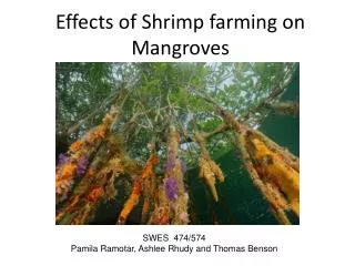
During the past 15 years, shrimp farming, mainly based on local species such as tiger shrimp<br>Penaeus monodon, kuruma shrimp Marsupenaeus japonicas and the exotic species white shrimp<br>Litopenaeus vannamei, have been particularly badly hit by epidemics associated with viruses<br>and Vibrio bacteria like Vibrio alginolyticus and V. parahaemolyticus due to deteriorated<br>environment.

E N D
Available online www.jocpr.com Journal of Chemical and Pharmaceutical Research __________________________________________________ J. Chem. Pharm. Res., 2011, 3(5):501-513 ISSN No: 0975-7384 CODEN(USA): JCPRC5 Immunostimulatory effects of Cardiospermum halicacubum against Vibrio parahaemolyticus on tiger shrimp Penaeus monodon T. Rajasekar1, J. Usharani2, M. Sakthivel1 and B. Deivasigamani1* 1Faculty of Marine Sciences, CAS Marine Biology, Annamalai University, Parangipettai, Tamil Nadu, India 2Sri Sankara Arts and Science College, Enathur, Kanchipuram, Tamil Nadu, India ______________________________________________________________________________ ABSTRACT During the past 15 years, shrimp farming, mainly based on local species such as tiger shrimp Penaeus monodon, kuruma shrimp Marsupenaeus japonicas and the exotic species white shrimp Litopenaeus vannamei, have been particularly badly hit by epidemics associated with viruses and Vibrio bacteria like Vibrio alginolyticus and V. parahaemolyticus due to deteriorated environment. Shrimp lack an adaptive immune system and rely on innate immune responses against microbial invasion. Therefore, the health of shrimp and enhancement of its immunity are of primary concern. Immunostimulants are agents/factors that trigger the non-specific immune response and result in enhanced disease resistance. Several compounds have been reported to have immunostimulation properties. Many of these are derivatives or cellular components of bacterial, fungal or animal origin. Laminarin, barley, glucan, lactoferrin, levamisole, lipopolysaccharides, curdlan, scleroglucan, zymosan, inulin, chitosan, β-glucans, dextran, lentinan, krestin, saponins, herbal extracts, peptidoglycans and so forth, are some of the examples of immunostimulants used in shrimp/fish aquaculture. This present study was attempted to study the immunomostimulatory effects of plant extract from herbal plants on shrimp through fed diet. The plant extract was mixed with fed diet with different concentration and non-specific immune response and THC, Phenoloxidase, NBT and lysozyme activity were measured. After feed diet experiment shrimp were challenge with isolated Vibrio spp. pathogen and cumulative survival rate were calculated. Key words: Immunostimulatory, Cardiospermum halicacubum, Vibrio parahaemolyticus, Penaeus monodon. ______________________________________________________________________________ 501
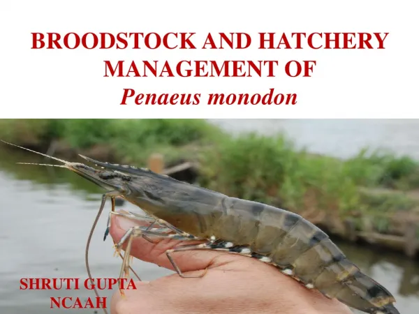
B. Deivasigamaniet al ______________________________________________________________________________ J. Chem. Pharm. Res., 2011, 3(5):501-513 INTRODUTION Shrimp culture is considered as one of the lucrative industries because of the high market price and great demand in the international market [1]. During the past 15 years, shrimp farming, mainly based on local species such as tiger shrimp Penaeus monodon, kuruma shrimp Marsupenaeus japonicas andthe exotic specieswhite shrimp Litopenaeus vannamei, have been particularly badly hit by epidemics associated with viruses [2]and Vibrio bacteria like Vibrio alginolyticus [3]and V. parahaemolyticus [4]due to deteriorated environment. Some of the insecticides and biocides used in aquaculture are known to accumulate and concentrate in aquatic organisms, while antibiotics may well induce resistance in pathogens through mutagenesis and plasmid mediated gene transfers [5].Evidence indicates that shrimp pathogens are opportunistic agents. Disease outbreaks are also associated with increases in the proportion of potentially pathogenic species in the Vibrio population of cultured pond waters [6]. Shrimp lack an adaptive immune system and rely on innate immune responses against microbial invasion [7]. Therefore, the health of shrimp and enhancement of its immunity are of primary concern. The innate immune system of shrimp consists mainly of prophenoloxidase (proPO) activating system, clotting system, phagocytosis, encapsulation and nodule formation, antimicrobial peptides (AMPs) formation and cell agglutination. The first and essential step of the internal defence process is the recognition of invading microorganisms, which is mediated by the pattern recognition proteins (PRPs). With antigenic components being recognized by PRPs, signaling cascades start and haemocytes become activated[8].The haemocytes are involved in the synthesis, storage and upon activation, discharge of proenzymes and substrates of the clotting and proPO system [9].The activated haemocytes are also induced to release other proteins that are related to defence responses, such as AMPs and peroxidase [10]. In order to control the proliferation of opportunistic bacteria, prophylactic chemo-therapeutants have been commonly used and practiced in intensive aquaculture. However the abuse of broad spectrum chemo-therapeutants has resulted in an increased number of antibiotic-resistant bacteria, the transfer of these drug-resistant genes to bacteria or virus infects terrestrial animals and humans, however alterations of the bacterial flora both in sediments and in water column and residual act as antibiotics in seafood products [11]. Therefore, it is necessary to exploit a non- chemotherapeutic method instead of the chemotherapeutic method, such as in the use of vaccine, probiotics, immunostimulants and natural therapeutics from plants [12]. Immunostimulants are agents/factors that trigger the non-specific immune response and result in enhanced disease resistance. Several compounds have been reported to have immunostimulation properties. Many of these are derivatives or cellular components of bacterial, fungal or animal origin. Laminarin, barley, glucan, lactoferrin, levamisole, lipopolysaccharides, curdlan, scleroglucan, zymosan, inulin, chitosan, β-glucans, dextran, lentinan, krestin, saponins, herbal extracts, peptidoglycans and so forth, are some of the examples of immunostimulants used in shrimp/fish aquaculture [13]. There are increasing number of investigations on plant extract activities of immune enhancement and anti-microorganism. Essential oils, methanol and ethanol 502
B. Deivasigamaniet al ______________________________________________________________________________ J. Chem. Pharm. Res., 2011, 3(5):501-513 extracts are known to possess antimicrobial activity [14]. Essential oils have been traditionally used for respiratory tract infections, and are used these days as ethical medicines for colds [15]. Now a days, the herbal drugs popularity increased and used widespread. The research is still lagging behind to get the efficacy of plant derived medicines on microorganisms which induced pathogenesis in Human beings and other animals. There were reports of various medicinal plants that had been used for years in daily life to treat disease all over the world. There were several reports on the antimicrobial activity of different herbal extracts in different regions of the world [16]. Phytomedicines derived from plants have shown great promise in the treatment of intractable infectious diseases [17]. A special feature of higher plants is their capacity to produce a large number of secondary metabolites [18].There was another aspect of drugs used against the microorganisms. The multiple drugs used against the microorganism had developed resistance due to the indiscriminate use of commercial antimicrobial drugs commonly used in the treatment of infectious disease [19]. Natural plant products have been reported to promote various activities like antistress, growth promotion, appetite stimulation, tonic and immunostimulation and to have aphrodisiac and antimicrobial properties in finfish and shrimp larviculture due to the active principles such as alkaloids, flavanoids, pigments, phenolics, terpenoids, steroids and essential oils [20][21]. This present study was attempted to study the immunomostimulatory effects of plant extract from herbal plants on shrimp through fed diet. The plant extract was mixed with fed diet with different concentration and non-specific immune response and THC, Phenoloxidase, NBT and lysozyme activity were measured. After feed diet experiment shrimp were challenge with isolated Vibrio spp. pathogen and cumulative survival rate were calculated. EXPERIMENTAL SECTION Plant materials In this present investigation plant leaf such Cardiospermum halicacubum (P4)was collected from Kanchipuram District, Tamil Nadu by using sterile polythene bags. Collected leaves were washed with distilled water and shadow dried. Preparation and preservation of plant extract The shadow dried plant leaves were grinded by using mixer grinder. Approximately 400 g of the plant materials were macerated with 80% methanol incubated for at room temperature and after 24 h filtered through Whatman filter paper No.1. The procedure was repeated three times to ensure exhaustive extraction of the plant material. The extracts were pooled together, concentrated and the solvent removed by evaporation under reduced pressure in a rotary evaporator at 40ºC []. Then extracts were dried by freeze-drying and kept in a freezer at −20ºC, until used for further studies. Feed preparation and experimental design To study the innate immune responses, four different concentration of plant extract C. halicacubum mixed with pellet feed based on the experiments were designed. Group 1: Commercial shrimp pellet (Control) Group 2: 0.1% of crude plant extract with commercial pellet 503
B. Deivasigamaniet al ______________________________________________________________________________ J. Chem. Pharm. Res., 2011, 3(5):501-513 Group 3: 0.5% of crude plant extract with commercial pellet Group 4: 1% of crude plant extract with commercial pellet Experimental diets were produced by combining commercial shrimp pellets with powdered cured plant extract and 50% gelatin (Knox) to produce four diets: (1) 0% Control commercial (containing 1% carrier ), (2) 0.1% , (3) 0.5%, (4) 1%. Dry components were ground to a powder using a mortar and pestle. Gelatin, dissolved at 60 µg ml-1 in boiling water, was cooled to 25°C and mixed at 50% v/w with the powdered diet. The resulting paste was spread evenly on aluminum foil and cooled at 4°C until solid. Pellets were then cut to approximately 0.5 cm3 cubes and stored in sealed plastic bags at 4°C for no longer than 7 days prior to use. Experimental shrimp Black tiger shrimp P. monodon (n=160) were obtained from a commercial farm in Sirkazhi area, Tamil Nadu, India and was acclimated to the laboratory condition for 5 days before the experimental period. During this period, shrimp was fed twice daily with a commercial pellet feed. Intermoult stage shrimp was used for the study. The moult stage was identified by examination of the uropoda in which partial retraction of the epidermis could be distinguished. All the experimental shrimp were morphologically identified for symptoms (shrimp reddishness, antenna rot, spots in carapace, gill coloration, wound and loose shells) and healthy shrimp were used for experiments. Shrimp ranged in weight from 16.5 to 19 g were used in these experiments. During the experiments, water (temperature, pH, salinity) conditions were observed as 20 – 28°C, 7.8- 8.0, and 22 – 24‰ respectively. The Immunostimulatory activity -Immunological parameters Haemolymph collection Haemolymph was collected with an insulin syringe with 28 gauge needle from the ventral region of the cephalothorax. Alsever’s solution (NaCl - 4.2 g-1, trisodium citrate- 8 g-1, citric acid 0.55 g ml-1, D-glucose 20.5g l-1) was used as an anticoagulant. Equal ratio of anticoagulant and haemolymph were mixed, from that 25 µl were used for THC (Total Haemocytes Count) analysis and NBT (Nitroblue tetrazolium) assay. Totally 100 µl were centrifuged at 10000 g for 10 min at 4°C and the supernatant was used to measure total phenoloxidase (PO) activity and lysozyme activity. Total haemocytes count Equally diluted haemolymph was further diluted with equal ratio of anticoagulant. Then, the THC in the diluted haemolymph were immediately determined using a Neubauer haemocytometer. THC on the heamocytometer were observed under Light microscope (Magnius MLX) and counted in all 4 squares by adaptation of routine method. Haemocyte count = N x D/A x 10 x 103 cells ml-1 (where, N = total number of cells counted; D = dilution of haemolymph; A = total area counted (in mm2); 10 = factor to convert area to volume (in µl), assuming a chamber of 0.1 mm depth; 103 = factor to convert count per µl to count per milliliter). 504
B. Deivasigamaniet al ______________________________________________________________________________ J. Chem. Pharm. Res., 2011, 3(5):501-513 Total phenoloxidase (PO) activity Total phenoloxidase activity was determined by using L-Dihydroxyphenylalanine (L-DOPA) (Perazzolo and Barracco, 1997). Briefly, 100 µl of centrifuged haemolymph was mixed with 50 µl PBS solution and 50 µl of enzyme inducer trypsin (Hi Media, 1 mg ml-1) and incubated for 15 min at 25°C in 96-microliter plates (flat bottomed). In controls, trypsin and serum were replaced by PBS. After incubation, 100 µl of a L-DOPA solution (10mg 1ml-1) were added to that mixture and incubated for 10 min at 25°C. Then the sample were read at 490nm using a VERSAmax tunable microplate reader (Associates of Cape Cod Incorporated, East Falmouth, MA, USA) [19]. Reduction of NBT by haemocytes To determine the amount of superoxide anion the reduction of nitroblue tetrazolium (NBT) by haemocytes (Respiratory burst activity) was measured [20]. Haemolymph (100 µl) was placed in a microplate and incubated for 30 min at room temperature. The supernatant was discarded and 50 µl of 0.3% NBT were added and incubated for 2 h at room temperature. The supernatant was again discarded, and the haemocytes were fixed with 200 µl of absolute ethanol. Haemocytes were washed twice with 200 µl 70% methanol and let dry. The formazan deposits generated were dissolved in 120 µl 2 M KOH and 140µl dimethyl sulfoxide (DMSO) and optical density at 620 nm was recorded using a microtiter plate reader (VERSAmax tunable microplate reader). Lysozyme activity Lysozyme activity was measured by the modified method of Minagawa [21]et al. In this turbidometric assay, 0.03% lyophilized Micrococcus lysodeikticus in PBS was used as substrate. Ten microlitres of haemolymph was added to 250 µl of bacterial suspension in duplicate wells of a microtitre plate and the reduction in absorbance at 490 nm was determined after every minutes (1, 2, 3, 4 and 5) of incubation at 22oC using a microtiter plate reader (VERSAmax tunable microplate reader). One unit of lysozyme activity was defined as a reduction in absorbance of 0.001 per min. Challenge with Vibrio parahaemolyticus In the present study, the isolated bacterial pathogen Vibrio parahaemolyticus (VP1) was used as a test organism. Lyophilized VP1 strain was used in this study .The bacterial strain VP1 was subcultured and centrifuged at 10000 g for 10 minutes at 4°C. The supernatant were discarded and the bacterial pellet was washed three times and resuspended in phosphate buffered saline (PBS) at pH 7.4. The OD of the solution was adjusted to 0.5 at 456 nm which corresponded to 1× 10⁷ cells ml-1. After plant extract treatment, shrimp were injected (50 µl) with V.parahaemolyticus (no 1) (1×10⁷ cells ml-1) on 6th day. RESULTS The Immunomodulatory activity-Immunological parameters Total haemocytes count After 24 h of experiment diet, slightly increased cell count of all experimental shrimp was observed except control. (Figure 1) After 48 h there was a gradual increase in cell count based on concentration wise and control showed normal cell count. Highest cell count was observed in G3 as 9.2 ×10⁷ cell ml-1 and lower was observed in G1 as 1.1 ×10⁷ cell ml-1. 505
B. Deivasigamaniet al ______________________________________________________________________________ J. Chem. Pharm. Res., 2011, 3(5):501-513 Total phenoloxidase (PO) activity There was a significant increase in group G2 and G3 after 24 h of fed diet experiment. The plant extraction concentration in fed diet there were a gradually increased in the entire three groups. After 48 h of fed diet maximum activity was observed in G3 group and minimum in G1 group (Figure 2). Reduction of NBT by haemocytes During the initial period and after 24 h of experiment diet, normal level of NBT (respiratory burst activity) activity were observed. After 48 h, NBT activity was gradually increased in all groups. After 72 h, G1 and G2 were significantly increased and G3 was slightly decreased. Maximum NBT activity was observed on 120 h of feed experiment in G3 group and minimum in G1 group. G1 and G3 showed similarly NBT activity when compared with control and G1 group (Figure 3). Lysozyme activity There is no significant change in lysozyme activity was observed in all experimental groups after 24 h. After 48 h, slight increase was observed in all groups (Figure 4). After 96 h significant increased was observed in all the experimental shrimp in response to fed diet concentration maximum lysozyme activity was observed in group G3 as 375 U ml-1 min-1 and minimum was observed in G1 as 340 U ml-1 min-1 . Cumulative survival rate After fed diet experiment shrimp were challenge with V.parahaemolyticus and observed for the survival rate and after 24h of challenge test, experiment group showed 90% survival and control 80% survival were observed .After 72 h 50 % of survival was observed in control. After 168 h of experimental studies 60% of survival was observed in G1 Group and 70% of survival was observed in G2 and G3, control shrimp 20% of survival was observed (Figure 5). DISCUSSION Crustaceans have only a non-specific innate immune response and no long memory, which results in the inability of shrimp to protect itself from infection with its immune system [22]. Earlier reports revealed that the extracted compounds were responsible for antibacterial activity which may be extracted by low polarity solvents rather than by solvents with high polarity such as water. Other alternative feed additives that can prevent disease and enhance growth have been used to replace drug such as antibiotic in aquaculture [12] [24]. Recently, the so-called natural and phytogenic extracts have gained interests in their utilization to improve immunity and disease resistance of farmed fish or shellfish. Immunostimulants are the substances that can enhance the non-specific defence mechanism in shrimp and provide resistance against pathogenic organism. Protective effects of various immunostimulants have been reported against WSSV infection, for example, oral administrationof peptidoglycan or lipopolysaccharide in Penaeus japonicus[25]and β 1–3 glucan in Penaeus monodon [26] [27]. Shrimp hemocyanin was also shown to exhibit antiviral activity against a variety of viruses including DNA and RNA viruses [28]. Mortality was reduced by a 506
B. Deivasigamaniet al ______________________________________________________________________________ J. Chem. Pharm. Res., 2011, 3(5):501-513 synthetic antibacterial peptide from Mytilus galloprovincialis in WSSV infected palaemonid shrimp[29]. Herbal immunostimulants from Cyanodon dactylon, Aegle marmelos, Tinospora cordifolia, Picrorhiza kurooa and Eclipta alba were found to be effective against WSSV [30]. The haemocyte count varies among crustacean species and is known to be affected by a variety of factors such as infection and environmental stress [31]. Persson [32] et al. demonstrated a decrease in the haemocyte numbers of crayfish harboring a parasitic fungus (Aphanomyces astaci). Maeda [33] et al. have observed a decline in total haemocyte count (THC) in shrimp infected with penaeid rod shaped DNA virus V. parahaemolyticus and V. harveyi also decreased the THC in white shrimp Litopenaeus vannamei [34]. Several studies have demonstrated that the use of immunostimulants alone [35] [36]lead to enhanced resistance to bacterial diseases in shrimp. In the present study, THC in experimental shrimp was estimated. In that, G3 group with 1% of plant extract fed shrimp showed maximum cell count and minimum was observed in G1 group. Secombes and Olivier [37]proposed that the release of superoxide anion, hydrogen peroxide, and hypochlorous acid (HOCl) into the phagosome and extracellular space during the respiratory burst is considered to be one of the most important mechanisms involved in the bactericidal activity of macrophages. Siwicki [38]et al. reported that oral administration of levamisole increased the phagocytic index of phagocytic cells. Additionally, it was reported that feeding yeast products (Macro Gard, Candida utilis, Saccharomyces cerevisiae) gave rise to an enhanced phagocytic activity of blood leukocytes in rainbow trout. A wide variety of standard dietary ingredients reported to affect immune responses. It is well known that fish and shellfish treated with immunostimulants showed increased phagocytosis as well as respiratory burst activity [39] [40] [41]. In this experiment after 48 h, NBT activity was gradually increased in all groups. After 72 h, G1 and G2 were significantly increased and G3 was slightly decreased. Maximum NBT activity was observed on 120 h of feed experiment in G3 group and minimum in G1 group. G1 and G3 showed similarly NBT activity when compared with control and G1. Since superoxide anion (O2-) is the first product to be released from the respiratory burst, the measurement of superoxide anion has been accepted as a precise way for measuring respiratory burst. Similar results in O2- production were observed for shrimp haemocytes stimulated with Zymosan[42] or with β- glucan[43]. The role of phenoloxidase in self-defense systems in sea cucumber may not be as critical as in crustaceans and insects. Lysosomal enzymes participate in the destruction of external substances in sea cucumber [44]. Regarding humoral defence mechanisms, the measurement of PO activity has been proposed as a procedure to evaluate the immune response of shrimps [45]. The activation of proPO system by microbial cell walls components such as β-1, 3-glucans from fungi and lipopolysaccharides (LPS) from Gram-negative bacteria has been demonstrated in several crustaceans species [46]. Lai [47] et al. stated that the phenoloxidase activity of L. vannamei was able to be activated by the leaf and twig hot-water extracts of C. kanekirae in agreement with the in vitro studies showed that laminarin and sodium alginate isolated from brown seaweeds, has been reported to 507
B. Deivasigamaniet al ______________________________________________________________________________ J. Chem. Pharm. Res., 2011, 3(5):501-513 increase the activity of the proPO system of penaeid shrimp[48]. It is considered that hot-water extracts of C. kanekirae can be used as an immunostimulant in shrimp diets for the improvement of shrimp immunity and disease resistance. Lysozyme is a fish defense element, which causes lysis of bacteria and activation of the complement system and phagocytes by acting as opsonin [49]. Elevated lysozyme activity was noted on 20, 25 and 30 days after feeding Jian carp [50]. and large yellow croaker, Pseudosciaena crocea [51] Astragalus root (Radix astragalin seu heydsari) and Chinese Angelica root (R. angelicae sinensis) at a ratio of 5:1 (w/w). In Oreochromis niloticus fed with 0.1 and 0.5% Astragalus radix root for 1 week, lysozyme activity was enhanced [52]. Dietary L-ascorbic administration at 288 mg kg-1 and dietary vitamin E administration at 100 mg kg-1 for 8 weeks have been reported to increase alternative complement and lysozyme activity of juvenile grouper Epinephelus malabaricus [53]. Sea bass Dicentrarchus labrax which had been fed a diet containing sodium alginate from brown algae Laminaria digitata and Ascofillum nodosum after 15 days showed increased alternative complement and lysozyme activity [54]. In the present study, After 48 h, exiguously increase was observed in all groups. After 96 h, significant increase was observed in all the experimental shrimp in response to fed diet concentration maximum lysozyme activity was observed in group G3 as 375 U ml-1 min-1 and minimum was observed in G1 as 340 U ml-1 min-1. Moreover the increased lysozyme activity can early produce good non specific immune response.In this present study, total phenoloxidase activity for 1% of P4 plant powder showed two fold activities than control shrimp. However moderate activity was observed in 0.5 and 1% of plant powder. Figure 1: Total haemocytes count with traditional Chinese medicine (TCM) formulated from 120 Contol G2 G1 G3 100 5cells/m l) 80 Total haem ocytes count (x10 60 40 20 0 -20 0 24 48 72 96 120 Time elapsed (h) Total haemocytes count of P. monodon during experimental feed. Results were made at initial 0 h, 24, 48, 72, 96 and 120h for different experimental feed composition (C, G1, G2 and G3). Each bar represents the mean value from three determinations with the standard deviation (SD). Mean ± SD. 508
B. Deivasigamaniet al ______________________________________________________________________________ J. Chem. Pharm. Res., 2011, 3(5):501-513 Figure 2: Total Phenoloxidase activity 0.32 Control G2 G1 G3 Total Phenoloxidase activity (OD. 490nm) 0.28 0.24 0.20 0.16 0.12 0.08 0.04 0.00 0 24 48 72 96 120 Time elapsed (h) Figure 3: Respiratory burst activity-NBT 0.20 Control G2 G1 G3 0.16 N B T activ ity (O D . 620n m ) 0.12 0.08 0.04 0.00 0 24 48 72 96 120 Time elapsed (h) 509
B. Deivasigamaniet al ______________________________________________________________________________ J. Chem. Pharm. Res., 2011, 3(5):501-513 Figure 4: Lysozyme activity Control G2 G1 G3 400 350 -1) -1m in 300 Lysozym e activity (U m l 250 200 150 100 50 0 0 24 48 72 96 120 Time elapsed (h) Figure 5: Cumulative survival rate Control G2 G1 G3 100 Cumulative survival rate (% ) 80 60 40 20 0 24 48 72 96 120 144 168 Time elapsed (h) CONCLUSION In this present study reveals the Cardiospermum halicacubum plant extract shows good immune stimulatory activity against shrimp and showed good survival rate after challenge with VP1. Further characterization and purification of plant extract and it could be a superior applicant for the development of good immune stimulatory agent in the aquaculture industry. 510
B. Deivasigamaniet al ______________________________________________________________________________ J. Chem. Pharm. Res., 2011, 3(5):501-513 Acknowledgement The authors are thankful to Professor Dr. T. Balasubramanian, Dean of our center for all support and facilities rendered and also sincere thanks to Principal, Sri Sankara Arts and Science College Kanchipuram , providing the lab facility during my project work. REFERENCES [1] Venkatramalingam K; Godwin C.J and Citarasu T. Aqua Nutri.,2007,13, 439-443. [2] Lo C.F; Chang Y.S; Peng S.E and Kou G.H. J. Fish Soc. Taiwan., 2003, 30, 1-13. [3] Liu C.H and Chen J.C. Fish and Shellfish Immunol., 2004, 16, 321-334. [4] Sung H.H; Hsu S.F; Chen C.K; Ting Y.Y and Chao W.L. Aquaculture, 2000,192, 101-110. [5] Panigrahi A and Azad I.S. Fish Physiology and Biochemistry, 2007, 33(4), 429-440 [6] Lavilla-Pitogo C.R; Leano E.M and Paner M.G. Aquaculture,1998, 164, 337-49 [7] Bulet P; Hetru C; Dimarcq J.L and Hoffmann. Dev. Comp. Immunol., 1999, 23, 329–344. [8] Vargas-Albores F and Yepiz-Plascencia G. Aquaculture, 2000, 191:13-21 [9] Sritunyalucksana K and Soderhall K. Aquaculture, 2000, 191, 53– 69. [10] Cerenius L and Soderhall K. Immunol. Rev., 2004, 198, 116-126. [11] Verschuere; Rombaut G; Sorgeloos P and Verstraete W. Microbiol. Mol. Biol. Rev., 2000, 64, 655-671. [12] Sakai M. Aquaculture, 1999, 172, 63-92. [13] Newman S.G, Deupree R.H, The impact of biotechnology on aquaculture. Aquaculture towards the 21st century. In: Proceedings of Infofish-Aquatech-94, International Conference on Aquaculture. Colombo, Sri Lanka, INFOFISH, Kuala Lumpur (Malaysia), 1994, 147–58 [14] Shahverdi A.R; Monsef-Esfahani H.R; Tavasoli F and Mirjani, R. J. Food Sci., 2007, 72, 55-58. [15] Federspil P; Wulkow R and Zimmermann T. Laryngorhinootologie, 1997, 76, 23-27. [16] Citarasu T; Sekar R.R; Babu M.M; Marian M.P. Developing Artemia enriched herbal diet for producing quality larvae in Penaeus monodon. Asian Fish Sci.,2002, 15, 21-32. [17] Sivaram V; Babu M.M; Citarasu T; Immanuel G; Murugadass S and Marian MP. Aquaculture, 2004, 237, 9-20. [18] Hamza J.M; Carolien J.P; Matee I.N; Moshi J; Mikxf H.M; Selemani O; Mbwambo H; Van der Ven J.A.M and Verweij E. J. Ethnopharmaco., 2006, 108, 124–132 [19] Mexia-Salazar A.L; Hernandez-Lopez J; Burgos-Hernadez A; Cortez-Rocha M.O; Castro- Longoria R and Ezquerr-Brauer J.M. Food chemistry., 2008, 110, 471-479. [20] Monoz M; Cedeno R; Rodriguez J; Vander W.P.W; Mialhe E and Bachere E. Aquaculture, 2000, 191: 89-107. [21] Maggioni D.S; Andreatta E.R; Hermes E.M and Barracco MA. Aquaculture, 2004, 241: 501-515. [22] Sarathi M; Ishaq A.V.P; Venkatesan C; Balasubramanian G; Prabavathy J and Sahul Hameed A.S. Aquaculture, 2007, 271, 8-20. [23] Sudheesh P.S and Xu H.S. Aquaculture, 2001, 196, 37-46. [24] Felix N; Vyla A.P and Jeyaseelan P. World Aqua., 2006, 37, 16-7. [25] Takahashi Y; Kondo M; Itami T; Honda T; Inagawa H and Nishiza T. Fish and Shellfish Immunol.,2000,10, 555-558. 511
B. Deivasigamaniet al ______________________________________________________________________________ J. Chem. Pharm. Res., 2011, 3(5):501-513 [26] Song YL, Liu JJ, Chan LC, Sung HH, Glucan-induced disease resistance in tiger shrimp (Penaeus monodon). In: Gudding, R., Lillehaug, A., Midtlyng, P.J., Brown, F. (Eds.), Fish Vaccinology, 1997, 413–421. [27] Chang C; Su M.S; Chen H.Y; Lo C.F; Kou G.H and Liao I.C. Dis. Aquat. Org., 1999, 36, 163–168. [28] Zhang X; Huang C; Tang X; Zhuang Y and Choy L.H. Proteins, 2004, 55, 229–235. [29] Dupuy J.W; Bonami J.R and Roch P.H J. Fish Dis., 2004, 27, 57–64. [30] Citarasu T; Sivaram V; Immanuel G and Rout N. Fish Shellfish Immunol., 2006, 21, 372– 384. [31] Smith V.J and Johnston P.A. Comp Biochem Physiol C., 1992, 101, 641–9. [32] Persson M; Cerenius L and Soderhall K. J Fish Dis., 1987, 10, 471–7. [33] Maeda M, Itami T, Kondo M, Henning O, Takahashi Y, Hirono I, et al. Characteristics of penaeied rod shaped DNA virus of kuruma shrimp. New approaches to viral disease of aquatic animals, NRIA international workshop, National Research Institute of Aquaculture. Nansei, Mie, Japan 1997:218–28. [34] Montero-Rocha D; McIntosh R; Sanchez-Merino I and Flores. J. of Invertebrate Pathology., 2006, 91: 188–194. [35] Itami T; Asano M; Tokushige K; Kubono K; Nakagawa A; Takeno N; Nishimura H; Maeda M; Kondo M; Takahashi Y. Aquaculture,1998, 164, 277–288. [36] Song YL, Hsieh YT, Immunostimulation of tiger shrimp (Penaeus monodon) haemocytes for generation of microbicidal substances: analysis of reactive oxygen species. Develop. Comp. Immunol., 1994, 18 (3), 201-209. [37] Secombes C.J and Olivier G. Academic Press, New York, 1997, 269-296. [38] Siwicki A.K; Anderson D.P; Dixon O.W. Develop. Comp. Immunol., 1990, 14: 231-237. [39] Siwicki A.K; Anderson D.P and Rumsey G.L. Vet. Immunol. Immunopathol.,1994, 41, 139-159. [40] Robertsen B, Engstad RE, Jorensen JB, Beta-glucans as immunostimulants in fish. In: Stolen, J.S., Fletcher, T.C, (Eds.). Modulators of fish immune responses: models for environmental toxicology, biomarkers, and immunostimulators. Fair Haven, NJ, USA: SOS Publications., 1994, 83-99. [41] Verlhac V; Obach A; Gabaudan J; Schuep W; Hole R. Fish and Shellfish Immunol., 1998. 8, 409-424. [42] Wang F.I. and Chen J.C. Aquaculture. 2006, 258: 34-41. [43] Song Y.L, Hsieh Y.T. Develop. Comp. Immunol., 1994, 18(3), 201-209. [44] Canicatti C. Invert. Pathol. 1990, 56, 70-74. [45] Rodriguez J and Moullac G.L. Aquaculture, 2000, 191, 109-119. [46] Suphantharika M; Khunrae P; Thanardkit P and Verduyn C. Bioresource Technol., 2003, 88, 55–60. [47] Lai, C.Y; Cheng W and Kuo C.M. Fish and Shellfish Immunol.,2005, 18, 417-30. [48] Hernandez-Lopez; Gollas-Galvan T and Vargas-Albores F. Comp. Biochem. Physiol., 1996, 113, 61-66. [49] Magnadottir, B. Fish and Shellfish Immuno., 2006. 20, 137–151. [50] Jian J.C and Wu Z.H. Fish and Shellfish Immuno., 2004,16, 185-91. [51] Jian J.C and Wu Z.H. Aquaculture. 2003, 218, 1-9. [52] Yin G; Jeney G; Racz T; Pao X; Jeney Z. Aquaculture, 2006, 253, 39-47. [53] Lin M.F and Shiau S.Y. Aquvaculture, 2005, 244, 215-221. 512
B. Deivasigamaniet al ______________________________________________________________________________ J. Chem. Pharm. Res., 2011, 3(5):501-513 [54] Bagni M; Romano N; Finoia M.G; Abelli L; Scapigliati G; Tiscar P.G and Sarti M; Marino G. Fish Shellfish Immunol., 2005, 18, 311–325. 513