1 / 112
1.12k likes | 1.35k Views
slideserve slideserve slideserve slideserve slideserve slideserve slideserve slideserve slideserve slideserve slideserve slideserve slideserve slideserve slideserve slideserve slideserve slideserve slideserve slideserve

E N D
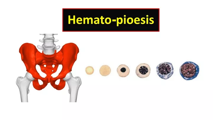
Hematopoietic Stem cell Myeloid progenitor Lymphoid progenitor
Lymphoid progenitor Small lymphocyte Natural killer cell (Large granular lymphocyte) B lymphocyte T lymphocyte Plasma cell
Myeloid progenitor Myelo-blast Pro-erythro-blast Mega-karyo-blast Monocyte Eosinophil Basophil Neutrophil Thrombocytes • (Platelet) Erythrocyte (RBCs)
Hematopoietic Stem cell • Lymphoid progenitor • Myeloid progenitor Lymho-blast Mono-blast Myelo-blast Pro-erythro-blast Mega-karyo-blast Small lymphocyte Large lymphocyte Monocyte Eosinophil Basophil Neutrophil Thrombocytes • (Platelet) Erythrocyte (RBCs) Granulocytes
1 2 3 4 Blood Cells Maturation
Blood Cell Maturation • A, Cell diameter decreases and cytoplasm becomes less basophilic. • • An exception to the diameter decreasing is observed in the granulocytic series • • In the erythroid series, hemoglobin development in the cytoplasm imparts a pink/salmon color. • B, Nuclear diameter decreases (N:C ratio decreases) • •Nuclear color changes from purplish red to dark blue. • C, Nuclear chromatin becomes coarser, clumped, and condensed. • • Nucleoli disappear. • • In the granulocytic series, the nuclear shape changes and the nucleus becomes segmented. Granules appear in cytoplasm • • In the erythroid series, the nucleus becomes fully condensed and is ejected. • D, Composite of changes during maturation process.
(Pro-erythro-blast) Pro-normo-blast Basophilic normoblast P R E C U Polychromatic normoblast R S O R Orthochromic normoblast P Polychromatic erythrocyte (reticulocyte) E R B I L P O H O E D Erythrocyte R A L
1. Pro-normo-blast SIZE: 12 to 20 μm Very Round cell NUCLEUS: Round to slightly oval Nucleoli: 1 to 2 Chromatin: Fine centrally-located nucleus nuclear pores CYTOPLASM: Dark blue; Basophilic may have prominent Golgi N:C RATIO: 8:1 REFERENCE INTERVAL: Bone Marrow: 1% , Peripheral Blood: 0% Nucleolus Cytoplasm Golgi Nucleus
Nucleolus Cytoplasm Golgi Nucleus
Pronormoblast Basophilic normoblast P R E C U Polychromatic normoblast R S O R Orthochromic normoblast P Polychromatic erythrocyte (reticulocyte) E R B I L P O H O E D Erythrocyte R A L
2. Baso-philic-normo-blast SIZE: 10 to 15 μm NUCLEUS: Round to slightly oval May binucleate Nucleoli: 0 to 1 Chromatin: Slightly condensed CYTOPLASM: Dark blue More basophilic N:C RATIO: 6:1 REFERENCE INTERVAL: Bone Marrow: 1% to 4% Peripheral Blood: 0% Cytoplasm Nucleus
Cytoplasm Nucleus
Pronormoblast Basophilic normoblast P R E C U Polychromatic normoblast R S O R Orthochromic normoblast P Polychromatic erythrocyte (reticulocyte) E R B I L P O H O E D Erythrocyte R A L
3. POLY-CHROMATIC -NORMOBLAST Cytoplasm SIZE: 10 to 12 μm NUCLEUS: Round Nucleoli: None Chromatin: Quite condensed A lot of nuclear pores CYTOPLASM: Gray-blue as a result of hemoglobinization N:C RATIO: 4:1 REFERENCE INTERVAL: Bone Marrow: 10% to 20% Peripheral Blood: 0% Nucleus
Cytoplasm Nucleus
Pronormoblast Basophilic normoblast P R E C U Polychromatic normoblast R S O R Orthochromic normoblast P Polychromatic erythrocyte (reticulocyte) E R B I L P O H O E D Erythrocyte R A L
4. Ortho-chromic-normoblast Cytoplasm SIZE: 8 to 10 μm NUCLEUS: Round Nucleoli: 0 Chromatin: Fully condensed CYTOPLASM: More pink or salmon than blue fullyhemoglobinized N:C RATIO: 0.5:1 About half is cytoplasm REFERENCE INTERVAL: Bone Marrow: 5% to 10% Peripheral Blood: 0% Nucleus
Cytoplasm Nucleus
Pronormoblast Basophilic normoblast P R E C U Polychromatic normoblast R S O R Orthochromic normoblast P Polychromatic erythrocyte (reticulocyte) E R B I L P O H O E D Erythrocyte R A L
5. Poly-chromatic-erythrocyte (Reticlocyte) SIZE: 8 to 8.5 μm NUCLEUS: Absent Nucleoli: NA Chromatin: NA CYTOPLASM: Color is slightly more blue/purple than the mature erythrocyte N:C RATIO: NA REFERENCE INTERVAL: Bone Marrow: 1% Peripheral Blood: 0.5% to 2.0%
Pronormoblast Basophilic normoblast P R E C U Polychromatic normoblast R S O R Orthochromic normoblast P Polychromatic erythrocyte (reticulocyte) E R B I L P O H O E D Erythrocyte R A L
6. Erythrocyte SIZE: 7 to 8 μm NUCLEUS: Absent Nucleoli: NA Chromatin: NA CYTOPLASM: Salmon with central pallor of about one-third of the diameter of the cell N:C RATIO: NA REFERENCE INTERVAL: Bone Marrow: NA Peripheral Blood: Predominant cell type
2. Granulocyte Maturation Neutrophil Basophil Eosinophil
Myeloblast Promyelocyte P R E C U Myelocyte R S O R Metamyelocyte P Band neutrophil E R B I L P O H O E Segmented neutrophil D R A L http://basicbook.net
1. Myeloblast SIZE: 15 to 20 μm NUCLEUS: Round to oval Nucleoli: 2 to 5 Chromatin: Fine typically has an eccentric nucleus. CYTOPLASM: Moderate basophilia Granules: Absent or up to 20 N:C RATIO: 4:1 REFERENCE INTERVAL: Bone Marrow: 0% to 2% Peripheral Blood: 0% Cytoplasm Nucleoli Nucleus
Cytoplasm Nucleoli Nucleus
Myeloblast Promyelocyte P R E C U Myelocyte R S O R Metamyelocyte P Band neutrophil E R B I L P O H O E Segmented neutrophil D R A L http://basicbook.net
2. Pro-myelocyte Cytoplasm with primary granules SIZE: 14 to 24 μm (slightly larger than myeloblast) NUCLEUS: Round to oval Nucleoli: 1 to 3 or more Chromatin: Fine, but slightly coarser than myeloblast CYTOPLASM: Basophilic , more a bundantGranules: PRIMARY: Granules more than 20; may be numerous; Red to purple or burgundy SECONDARY: None N:C RATIO: 3:1 REFERENCE INTERVAL: Bone Marrow: 2% to 5% Peripheral Blood: 0% Nucleus Nucleoli
Cytoplasm with primary granules Nucleus Nucleoli
Myeloblast Promyelocyte P R E C U Myelocyte R S O R Metamyelocyte P Band neutrophil E R B I L P O H O E Segmented neutrophil D R A L http://basicbook.net
3. Myelocyte Nucleus SIZE: 12 to 18 μm NUCLEUS: Round to oval; slightly eccentric; may have one flatten side; may have a clearing next to the nucleus indicating the location of the Golgi Nucleoli: Usually not visible Chromatin: Coarse and more condensed than promyelocyte CYTOPLASM: Slightly basophilic, to cream-colored, more a bundant Granules: PRIMARY: Few to moderate SECONDARY: Variable number; becoming predominant as cell matures N:C RATIO: 2:1 REFERENCE INTERVAL: Bone Marrow: 5% to 19% Peripheral Blood: 0% Golgi Cytoplasm with primaryand secondarygranules
Nucleus Golgi Cytoplasm with primaryand secondarygranules
Myeloblast Promyelocyte P R E C U Myelocyte R S O R Metamyelocyte P Band neutrophil E R B I L P O H O E Segmented neutrophil D R A L http://basicbook.net
4. Metamyelocyte SIZE: 10 to 15 μm NUCLEUS: Indented; kidney bean shape; indentation is less than 50% of the width of a hypothetical round nucleus Nucleoli: Not visible Chromatin: Moderately clumped CYTOPLASM: Pale pink, to cream colored, to colorless Granules: PRIMARY: Few SECONDARY: Many (full complement) N:C RATIO: 1.5:1 Nucleus 1.5 more than cytoplasm or equal REFERENCE INTERVAL: Bone Marrow: 13% to 22% Peripheral Blood: 0% Cytoplasm Nucleus
Myeloblast Promyelocyte P R E C U Myelocyte R S O R Metamyelocyte P Band neutrophil E R B I L P O H O E Segmented neutrophil D R A L http://basicbook.net
5. Band neutrophil Cytoplasm SIZE: 10 to 15 μm NUCLEUS: Constricted but no threadlike filament; indentation is more than 50% of the width of a hypothetical round nucleus NOTE: Chromatin must be visible in constriction; may be folded over. Nucleoli: Not visible Chromatin: Coarse, clumped CYTOPLASM: Pale pink, to colorless Granules: PRIMARY: Few SECONDARY: Abundant N:C RATIO: Cytoplasm predominates REFERENCE INTERVAL: Bone Marrow: 17% to 33% Peripheral Blood: 0% to 5% Nucleus
Cytoplasm Nucleus
Myeloblast Promyelocyte P R E C U Myelocyte R S O R Metamyelocyte P Band neutrophil E R B I L P O H O E Segmented neutrophil D R A L
6. Neutrophil SIZE: 10 to 15 μm NUCLEUS: 2 to 5 lobes connected by thin filaments, without visible chromatin Nucleoli: Not visible Chromatin: Coarse, clumped CYTOPLASM: Pale pink, cream-colored,or colorless Granules: PRIMARY: Rare SECONDARY: Abundant N:C RATIO: Cytoplasm predominates REFERENCE INTERVAL: Bone Marrow: 3% to 11% Peripheral Blood: 50% to 70% Cytoplasm Nucleus
Cytoplasm Nucleus