Download
1 / 22
290 likes | 1.14k Views
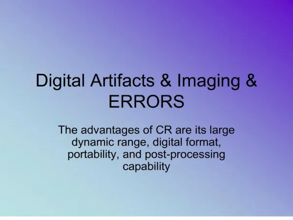
Digital Artifacts. Edge Enhancement. High contrast and edge definition Lose fine detail on the edges of high contrast areas. Ghost Image. CR Ghost Images.

E N D
Edge Enhancement • High contrast and edge definition • Lose fine detail on the edges of high contrast areas
CR Ghost Images Ghost or memory artifacts occur when the radiation remains trapped in the phosphor plate. Remember, radiation can remain trapped for several minutes. CR plates must be correctly erased to prevent ghost images.
Main Menu Key Advantages of Envision’s CMOS Line Array Technology The "fill factor" of a detector is the percentage of the surface area that is actually active, that is, capable of detecting photons. For example, where Envision's CMOS detector arrays have a fill factor in excess of 90%, the amorphous silicon detectors have a fill factor of only about 63%. The higher the fill factor, the more sensitive the detector is. 6. Fill Factor
Dead Pixels Dead PixelsThe number of known dead (nonfunctional) pixels and pixel aggregations was obtained from the system calibration table (bad-pixel mask). Calibration algorithms detect individual pixels, pixel clusters, and lines of contiguous pixels (rows or columns) that fail to produce a usable output value. In routine clinical use, a proprietary correction algorithm is applied to each image at the site of each nonfunctional pixel to fill in the missing image information on the basis of surrounding image information.
Printer Distortion • Occurs when the image size and the printed size are not equal.
Moire Pattern Grid lines run in the same direction as the laser in the CR reader.
The functionality of the RIS reflects the entire administrative workflow within the radiology department (see below), including: 1. Communication: Examination requests are interfaced from the HIS. Verified reports are interfaced back to the HIS, including billing information. 2. Scheduling: Examination requests are planned for a certain date, time, and modality, considering availability of resources. 3. Examination: Documentation of the examination performed, and additional data such as contrast media used, exposure dose, complications, etc. 4. Reporting: Assists the reporting radiologist by providing information not readily available in the PACS (eg clinical data, examination request forms). Facilitation of report transcription and verification. 5. Analysis: Management of cases for scientific work, statistical analysis, and documentation (eg the