Download
1 / 17
170 likes | 402 Views
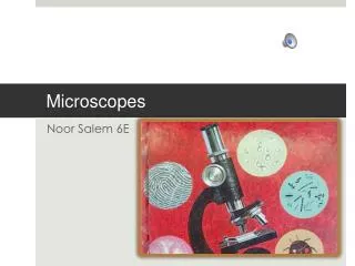
Microscopes. Microscopes are instruments used to produce an enlarged image of an object. When dealing with microscopes there are 2 very Important terms!. Magnification. Resolution. Why???. Which is more important??. An increase in an object’s apparent size.

E N D
Microscopes are instruments used to produce an enlarged image of an object. When dealing with microscopes there are 2 very Important terms! Magnification Resolution Why??? Which is more important?? An increase in an object’s apparent size. The amount of detail in the image.
Compound Light Microscope • In this class, we use a compound light microscope. • It uses light and two lenses (compound) to produce an enlarged image. • Light must pass THROUGH the specimen, so your specimen must be thin!
This is the microscope you’ll use in this classroom. OCULAR LENS – Has a magnification of 10x. This is the lens you look through. Draw it!! BODY TUBE – Connects ocular lens to the objective lenses. You raise this up and down to focus. NOSEPIECE – Holds the objective lenses. COARSE ADJUSTMENT KNOB – Moves the body tube up and down a lot. (yes really) FINE ADJUSTMENT KNOB – Moves the body tube up and down a little. ARM – Connects the body tube to the base of the microscope. STAGE – Place where you set your specimen. Has a hole through which the light passes. Why do we need light again?? PIVOT JOINT – Enables microscope to tilt. Don’t do it! Your slide might fall. BASE – Provides stability and support to the microscope.
SCANNING POWER – Has a magnification of 4x. You use this first to get an overview of the slide you’re viewing. Here is the nosepiece again. It rotates! STAGE CLIPS – These hold the slide in place. Handy if you’re not good at delicate movements! A good rule of thumb: the larger the objective lens…. the higher the magnification! HIGH POWER – Has a magnification of 40x. This is the last lens you will use. It is the most difficult! LOW POWER – Has a magnification of 10x. This is the second lens you will use when looking at a slide. Above you can see the 3 objective lenses. These enable you to increase/decrease the magnification of the object. Again, here is the stage … you can see the hole for the light!
DIAPHRAGM – Controls the amount of light passing through the stage. LIGHT SOURCE – Provides the light! Back in the day, this used to be a mirror!!!
Here you can see the diaphragm again. Notice the different sized holes. You will want to play around to find the best amount of light for each slide! The higher the magnification the more light you will need!
Calculating Total Magnification • Because these are COMPOUND microscopes and use 2 lenses, we need to multiply the magnification of the 2 lenses together. • You will always be using the ocular lens which has a magnification of 10x. • You will multiply the magnification of the objective lens you are using by 10x (the magnification of the ocular lens.
Let’s Try It!!!! If you are viewing an object in low power… What is the total magnification of that object? Calculate the magnification of high power!!!! If you are viewing an object in scanning power… What is the total magnification of that object? Low power is 10x. Ocular lens is 10x. So… 10 x 10 = 100x Scanning power is 4x. Ocular lens is 10x. So… 4 x 10 = 40x
How to Use the Microscope • Start with the body tube all the way down. • Set the slide on the stage so that the part of the slide you want to view is over the hole. • Make sure you are on scanning power. • Look through the ocular and slowly raise the body tube using the coarse adjustment.
Using the Microscope cont… • When you have the image in focus, observe, draw, move the slide around etc. • When you are ready, rotate the nosepiece to move the low power lens into place. • The image should need just a BIT of focusing. Use the coarse and fine adjustment to do so. • When you have the image in focus, observe, draw, move the slide around etc.
Using the Microscope cont.. • When you are ready for high power, rotate the nosepiece so the high power objective is in place. • Using FINE ADJUSTMENT ONLY, focus the image. • When you have the image in focus, observe, draw, move the slide around etc. • When finished, rotate the nosepiece to put the scanning power objective in place again.
MAJOR RULES • ALWAYS: Carry the microscope with 2 hands. One on the arm and one on the base. • NEVER: Use the coarse adjustment in high power. You might jam the objective into the slide and break it! • DON’T: Move the adjustment knobs before you switch between powers, you don’t have to! • NEVER: Clean the lenses! Let me do it for you! You cannot ever use water!
Drawing what you see under the microscope. Your field of vision in a microscope is circular so what you draw should be inside of a circle! Draw what you see. Draw objects where you see them! Label your drawings! Name of object. Magnification Half Moons 100x
Electron Microscope! Other Microscopes • The compound light microscope is just one type of microscope used in science. • Electron microscopes use a beam of electrons instead of light. They can magnify things up to 200,000x!! • Electron microscopes are large and expensive, but you can get some really neat images! White Blood Cell Butterfly Tongue! Spider! Bird Feather 1500x
Other Microscopes cont.. • You may also encounter a dissecting microscope. • These are used to view specimens through which light cannot pass. • They are basically a powerful magnifying glass!
Don’t wait! Ask Questions!