Download

1 / 1
10 likes | 134 Views
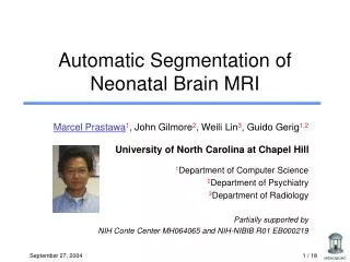
#. wm. gm. wm myl. Int. white. gray. csf. myelin. Template MRI. white matter. gray matter. csf. Here Text Here Text. Tissue Probability Maps. Assessing Early Brain Development in Neonates by Segmentation of High-Resolution 3T MRI

E N D
# wm gm wm myl Int white gray csf myelin. Template MRI white matter gray matter csf Here Text Here Text Tissue Probability Maps Assessing Early Brain Development in Neonates by Segmentation of High-Resolution 3T MRI 1,2G Gerig, 2M Prastawa,3W Lin, 1John Gilmore Departments of 1Psychiatry, 2Computer Science, 3Radiology University of North Carolina, Chapel Hill,NC 27614, USA gerig@cs.unc.edu / http://www.cs.unc.edu/~gerig RESULTS SUMMARY T1-only segmentation • Research: Quantitative MRI to study unsedated newborns at risk for neurodevelopmental disorders. • Clinical Study: 120 newborns recruited at UNC, age at MRI about 2 weeks • Motivation: Early detection of abnormalities Possibility for early intervention and therapy. • Imaging: High field (3T Siemens Allegra), high resolution (T1 1mm3, FSE 0.9x0.9x3mm3), high-speed imaging (12’ for T1, FSE and DTI). hyper- intense motor cortex early myelinated corticospinal tract Adult Neonate • Challenge: Very low CNR, heterogeneous tissue, early myelination regions, reverse contrast wm/gm. • Standard brain tissue segmentation fails. PD/T2 segmentation T1 3D MPRage 1x1x1 mm3 FSE T2w 1x1x3 mm3 FSE PDw 1x1x3 mm3 METHODS • Approach: • Atlas-moderated EM segmentation (cf. Leemput and Warfield) • Tissue intensity model for white matter (non-myelinated and myelinated wm form bimodal distribution) (cf. Cocosco, Prastawa) High-resolution PD/T2 data courtesy of Petra Hueppi, Univ. of Geneva. Preliminary Results UNC Neonate Study • So far: 20 normal neonates (10 males, 10 females) • Age 16 ± 4 days • Siemens 3T head-only scanner • Neonates were fed prior to scanning, swaddled, fitted with ear protection and had their heads fixed in a vac-fix device • A pulse oximeter was monitored by a physician or research nurse • Most neonates slept during the scan • Motion-free scans in 13-15 infants Building of Atlas Template CONCLUSIONS • It is feasible to study brain development in unsedated newborns using 3T MRI • Study will likely provide a vastly improved understanding of early brain development and its relationship to neuropsychiatric disorders. • Novelty: Tissue model for segmentation of myelinated/nonmyel. white matter. Literature • Gilmore JH, Gerig G, Specter B, Charles HC, Wilber JS, Hertzberg BS, Kliewer MA (2001a): Neonatal cerebral ventricle volume: a comparison of 3D ultrasound and magnetic resonance imaging. Ultrasound Med and Biol 27:1143-1146. • Huppi PS, Warfield S, Kikinis R, Barnes PD, Zientara GP, Jolesz FA, Tsuji MK, Volpe JJ (1998b): Quantitative magnetic resonance imaging of brain development in premature and normal newborns. Ann Neurol 43: 224-235. • Zhai G, Lin W, Wilber K, Gerig G, Gilmore JH (2003): Comparisons of regional white matter fractional anisotrophy in healthy neonates and adults using a 3T head-only scanner. Radiology (in press). • Warfield, S., Kaus, M., Jolesz, F., Kikinis, R.: Adaptive template moderated spa-tially varying statistical classification. In Wells, W.M.e.a., ed.: Medical Image Computing and Computer-Assisted Intervention (MICCAI’98). Volume 1496 of LNCS., Springer 1998 • Van Leemput, K., Maes, F., Vandermeulen, D., Suetens, P.: Automated model-based tissue classification of MR images of the brain. IEEE Transactions on Medical Imaging 18 (1999) 897–908 • Cocosco, C.A., Zijdenbos, A.P., Evans, A.C.: Automatic generation of training data for brain tissue classification from mri. In Dohi, T., Kikinis, R., eds.: Medical Image Computing and Computer-Assisted Intervention MICCAI 2002. Volume 2488 of LNCS., Springer Verlag (2002) 516–523 • Prastawa, M., Bullitt, E., Gerig, G., Robust Estimation for Brain Tumor Segmentation, MICCIA 2003, Nov. 2003 Supported by NIH Conte Center MH064065, Neurodevelopmental Disorders Research Center HD 03110 and the Theodore and Vada Stanley Foundation MICCAI Nov. 2003