Download
1 / 19
190 likes | 328 Views
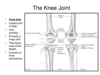
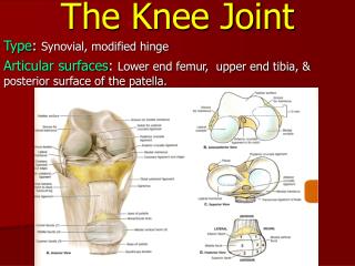
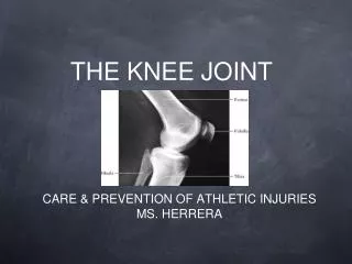
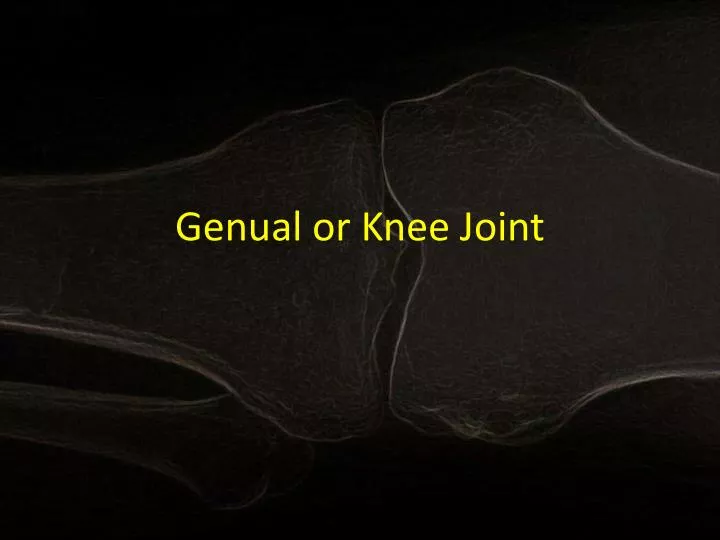
Genual or Knee Joint. Basic Information. Bones: Femur, Patella, and Tibia Location: 3 articulations; between condlyes of femur and tibia ( tibiofemoral joint) as well as between the patella and femur ( patellofemoral joint) Type of joint:

E N D
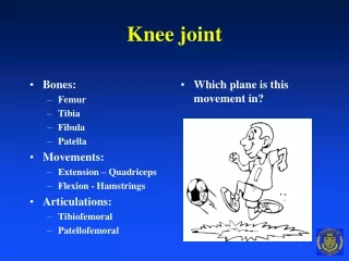
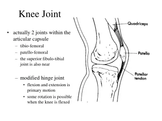
Basic Information • Bones: Femur, Patella, and Tibia • Location: 3 articulations; between condlyes of femur and tibia (tibiofemoral joint) as well as between the patella and femur (patellofemoral joint) • Type of joint: • Tibiofemoral-bicondylar/condyloid (modified hinge) • Patellofemoral- planar or gliding • Planes of Motion: • Tibiofemoral- biaxial (uniaxial with some rotary movement • Patellofemoral- uniaxial
Motion Permitted and Muscles that Permit Motion • Flexion • Gracillis • Sartorius • Biceps femoris • Semitendonosis • Semimembranosus • Popliteus • Gastrocnemius • plantaris
Motion Permitted and Muscles that Permit Motion (cont.) • Extension • Tensor fasciae lata • Rectus femoris • Vastuslateralis • Vastusmedialis • Vastusintermedius
Motion Permitted and Muscles that Permit Motion (cont.) • Medial Rotation • Gracilis • Sartorius • Semitendinosus • Semimembranosus • Popliteus
Motion Permitted and Muscles that Permit Motion (cont.) • Lateral Rotation • Tensor fascia lata • Biceps femoris
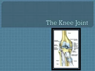
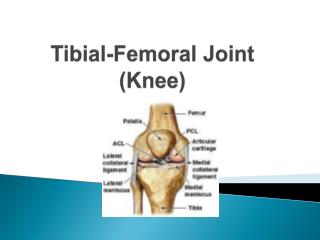
Ligaments • Major Ligaments
Important Structures • Bursae: Anteriorly there is the prepatellar, suprapatellar, and deep and superficial infrapatellar. • The prepatellar lies in the subcutaneous tissue between the skin and patella. The sac of the bursa is usually very thin, but becomes thick and distinct as the result of irritation; It is often enlarged, constituting "housemaid's knee”. The prepatellarbursae also allow the skin to move freely during movements of the knee. • The suprapatellar extends about two to three inches above the patella beneath the crureus muscle and is liable to be affected by stab or puncture wounds which would cause an infection in the joint. The suprapatellar bursa communicates freely with the synovial cavity of the knee. • The infrapatellar bursa are one between the skin and tibial tubercle and the other between the under surface of the tendo patellae and the upper end of the tibia and is unconnected with the joint and is not often diseased. The infrapatellarbursae also allow the skin to move freely during movements of the knee.
Important Structure (cont.) • Popliteus muscle: the tendons of this muscle separate the lateral meniscus from the LCL. It also helps with unlocking the knee when the leg is extended.
Important Structures (cont.) Articular capsule: - Encloses the posterior and lateral parts of the knee joint. It is supported by the LCL and MCL. It contains the patella, ligaments, menisci, and bursae. The capsule consists of a synovial and a fibrous membrane separated by fatty deposits anteriorly and posteriorly.
Important Structures (cont.) • The Meniscus -Located between the femur and tibia -C-shaped piece of fibrocartilage -Has no blood supply • Roles of the Meniscus -Shock absorption – absorbs the shock of the body weight -Stability – prevents the femur from rocking side to side on the tibia -Lubrication- prevents the friction between bones
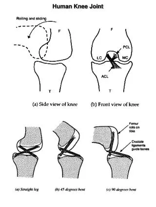
Additional Notes • The Knee joint is the largest and most complex diathroidal joint • Femur slants medially at the knee, and the tibia is vertical • Relatively weak mechanically • Stability is dependent on strength of ligaments and actions surrounding the muscle
Common Injuries • Sports Injuries Video • Sports Injuries 2 • The ACL stabalizes and aids in roatiation within the knee • Common injuries such as a pull or tear occur when foot is planted while the person is trying to pivot. This stretches the ACL reuslting in a pull or tear depending on the degree of stretching • The MCL's main function is to prevent the leg from extending too far inward, but it also helps keep the knee stable and allows it to rotate. • Comon injuries occur when outside of knee is hit which results in the MCL stretching to the point where it pulls or tears depending on the degree
Common Injuries (cont.) • Meniscal Injuries • -these types of injuries are extremely common among athletes as well as very active people. These injuries occur most when the knee is bent and then twisted. • Symptoms • -Knee pain • -Swelling of the knee • -Tenderness when pressing on the meniscus • -Popping or clicking within the knee • -Limited motion of the knee joint
Genetic Disorders/Diseases • Patellofemoral Syndrome • Osgood-Schlatter’s Disease • Jumper’s Knee (Petallartedinopathy) • Housemaid’s Knee • Osteoarthritis • Rheumatoid Arthritis
Prevention • KEEP YOUR FEET AND KNEES ALIGNED IN A STRAIGHT LINE • Whenever you are doing an exercise that requires bending, it is VERY IMPORTANT to make sure that your knees are aligned with your feet. This is VERY important if you are doing lunges or you are squatting. If your feet are not aligned with your knees, you will be putting too much stress on the knee joint and it could cause damage. • HAVE GOOD SHOES • Having a good cushion is extremely important for your joints. Spend the money to avoid injuries! • INCREASE PELVIC & THIGH MUSCLE STRENGTH • Most of the time, the knees pick up slack from the rest of our legs. If you increase the pelvic and thigh muscle strength, you will shift the balance back to where it should be. It reduces your knees’ workloads and decreases chances of being injured. • WARM UP & STRETCH • Warm up your legs and STRETCH before any exercise. This greatly reduces the risk for injuries. • NEVER OVERDO IT • Don’t strain your knee joints with workouts that aren’t realistic. Not everyone can bench 200 pounds or run 10 miles so work up to these goals, SLOWLY • BE KNOWLEDGABLE • If you already have BAD KNEES, try low impact workouts such as swimming or yoga. DON’T PUT TOO MUCH STRAIN ON YOUR KNEES!
Work Cited • http://www.arthroscopy.com/sp05001.htm • http://www.youtube.com/watch?v=95ZL6cv9Ikk • http://www.totaljoints.info/knee_joint_diseases.htm • http://www.youtube.com/watch?v=MIAr59bCs0A