Download
1 / 1
10 likes | 140 Views
The granule cell input - Purkinje cell output function. Introduction. Cerebellar dysfunction induces dystonia.

E N D
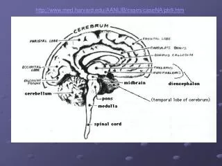
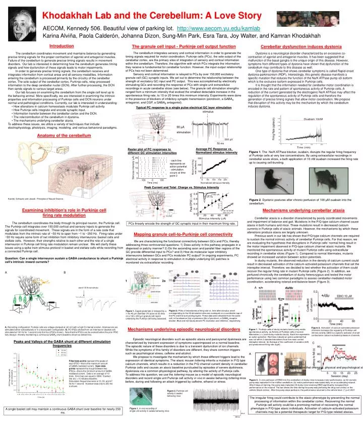
The granule cell input - Purkinje cell output function Introduction Cerebellar dysfunction induces dystonia • The cerebellum coordinates movement and maintains balance by generating precise timing signals for the proper contraction of agonist and antagonist muscles. Failure of the cerebellum to generate precise timing signals results in movement disorders. Our lab is interested in determining how the cerebellum generates timing signals and how dysfunction of these signals leads to motor impairments. • In order to generate precise timing signals, the cerebellum receives and integrates information from cortical areas and all sensory modalities. Information entering the cerebellum is processed primarily by the circuitry of the cerebellar cortex. The sole output of the cerebellar cortex, Purkinje cells, relay processed information to the deep cerebellar nuclei (DCN). After further processing, the DCN then sends signals to various target areas. • Our lab focuses on examining the cerebellum from the single cell level up to the behaving animal. More specifically, we are interested in examining the intrinsic properties and information processing of Purkinje cells and DCN neurons under normal and pathological conditions. Currently, our lab is interested in understanding: • How alterations in calcium homeostasis modulate Purkinje cell activity. • How Purkinje cells integrate and encode synaptic input. • Information transfer between the cerebellar cortex and the DCN. • The role/contribution of the cerebellum in dystonia. • The mechanisms underlying cerebellar ataxia. • To address these issues, we utilize a variety of techniques that include electrophysiology, photolysis, imaging, modeling, and various behavioral paradigms. Dystonia is a neurological disorder characterized by an excessive co-contraction of agonist and antagonist muscles. It has been suggested that malfunction of the basal ganglia is the unique origin of this disease. However, symptoms from different types of dystonia have shown that dysfunction of the cerebellum may contribute to this disease as well. One type of dystonia that shows cerebellar symptoms is called Rapid-onset dystonia-parkinsonism (RDP). Interestingly, this genetic disease manifests a specific mutation that reduces the function of the Na/K ATPase pump α3 isoform which is the exclusive isoform expressed in Purkinje cells. It is thought that the information needed for cerebellar motor coordination is encoded in the rate and pattern of spontaneous activity of Purkinje cells. A reduction of the current generated by the electrogenic Na/K ATPase may affect the regulation of the spontaneous activity of Purkinje cells and therefore the generation of precise timing signals that allow motor coordination. We propose that disruption of this activity may be the mechanism by which the cerebellum induces dystonia. The cerebellum integrates sensory and cortical information in order to generate the timing signals required for motor coordination. Purkinje cells (PCs), the sole output of the cerebellar cortex, are the primary sites of integration of sensory and cortical information within the cerebellum. Therefore, the algorithm with which PCs integrate the information they receive is fundamental for cerebellar function. However, the input-output relationship of PCs has not been determined. Sensory and cortical information is relayed to PCs by over 150,000 excitatory granule cell (GC) synaptic inputs. We set out to determine the relationship between the strength of excitatory GC input and PC output. This was accomplished by electrically stimulating GCs and recording the response of PCs with single-unit extracellular recordings in acute cerebellar slices (see below). The granule cell stimulation strengths ranged from a minimum intensity that evoked the smallest detectable increase in the spontaneous firing rate, to 10 or 20 times this minimum intensity. Experiments were done in the presence of blockers of inhibitory synaptic transmission (picrotoxin, a GABAA antagonist, and CGP, a GABAB antagonist). A B Figure 1. The Na/K ATPase blocker, ouabain, disrupts the regular firing frequency of Purkinje cells at very low concentrations. By using extracellular recordings in cerebellar acute slices, a bath application of 10 nM ouabain increased the firing rate up to causing cell-bursting. A B C Peak Current and Total Charge vs. Stimulus Intensity Kandel, Schwartz and Jessell. Principles of Neural Science. Figure 2. Dystonic postures after chronic perfusion of 100 µM ouabain into the cerebellum. PCs linearly encode the strength of GC synaptic input in their maximum firing rate. Figure 2. Activation of calcium-activated potassium channels increases the regularity of Purkinje cell intrinsic activity. EBIO is a specific activator of small-conductance calcium-activated potassium channels. Figure 1. Purkinje cells of ducky mutants have a very erratic spontaneous activity. (a) Activity of Purkinje cells of normal littermates (+/+) and ducky mutants (du/du). Each line represents an action potential. Red lines indicate an action potential that was not within 2 standard deviations from the mean control interspike interval. (b) Average of the coefficient of variation (left) and predominant firing rate (right). Figure 3. In vivo perfusion of EBIO into the cerebellum of ducky mice increases motor performance. (a) An osmotic pump was implanted in the midline cerebellum. (b) motor performance was tested daily on an accelerating rotarod. After 8 days of training, the pump was implanted. © ducky mice receiving EBIO significantly increased their performance on the rotarod. The bar shows the time during the pump was perfusing the drug (red circles) or the vehicle (black circles). After the pump stops perfusion, the performance returned to the vehicle level. (*) p<0.0001 The irregular firing could contribute to the ataxic phenotype by preventing the normal processing of information within the cerebellar cortex. Recovering the normal Purkinje cell activity could be a promising method of recovering the normal phenotype in P/Q-type ataxic individuals. Activation of calcium-activated potassium channels may be a potential therapeutic target for P/Q-type related ataxias. Khodakhah Lab and the Cerebellum: A Love Story Typical PC response to a single pulse electrical GC layer stimulation stimulus 90 µA stimulation Anatomy of the cerebellum 200 µV 50 ms Average PC Response vs. Normalized stimulus intensity Raster plot of PC responses to different GC stimulation intensities - each line represents an action potential - stimulation occurs at time point 0 Mechanisms underlying cerebellar ataxia Examining inhibition’s role in Purkinje cell firing rate modulation The cerebellum coordinates the body through its principal neuron, the Purkinje cell. The Purkinje cell integrates over 150,000 cortical and sensory inputs to generate the signals for coordinated movement. Those signals are in the form of a rate code that modulates from the intrinsic rate of ~50 Hz to span from ~1 to ~250 Hz. Firing rates under ~50 Hz require some form of net inhibition from inhibitory interneurons: basket cells and stellate cells. However, their strengths relative to each other and the role of a single interneuron in Purkinje cell firing rate modulation remain unclear. We will clarify these issues using a spike train stimulus protocol in basket and stellate cells while recording from a connected Purkinje cell. Question: Can a single interneuron sustain a GABA conductance to shunt a Purkinje cell’s intrinsic inward currents? A B Cerebellar ataxia is a disorder characterized by poorly coordinated movements and impairment of balance and gait. Mutations in the P/Q-type calcium channel cause ataxia in both humans and mice. These mutations result in decreased calcium currents in Purkinje cells of ataxic animals. However, the mechanisms by which these alterations produce ataxia are largely unknown. Previous work in our lab has shown that P/Q-type calcium channels are required to sustain the normal intrinsic activity of cerebellar Purkinje cells. For that reason, we are evaluating the hypothesis that disruptions in Purkinje cells’ normal firing result in the motor impairment observed in P/Q-type calcium channel ataxic mutants. We monitored the spontaneous activity of mutant Purkinje cells using extracellular recording in cerebellar slices (Figure 1). Compared to normal littermates, mutants showed an increased variation between action potentials. In ducky mutants, the observed reduction in the density of calcium current could result in decreased activation of the calcium-activated potassium channels that set the interspike interval. Therefore, we decided to test whether the activation of them could recover the regular firing rate in mutant Purkinje cells (Figure 2). In addition, we perfused chronically the cerebellum of ducky heterozygous and tested the motor performance using two common paradigms to assess cerebellar-mediated motor coordination, accelerating rotarod and balance beam (Figure 3). AECOM, Kennedy 506. Beautiful view of parking lot. http://www.aecom.yu.edu/kamlab Karina Alviña, Paola Calderón, Johanna Dizon, Sung-Min Park, Esra Tara, Joy Walter, and Kamran Khodakhah Mapping granule cell-to-Purkinje cell connectivity We are characterizing the functional connectivity between GCs and PCs, thereby addressing three controversial questions: 1) Does activity in this pathway propagate in a dispersed or patchy manner? 2) Do the ascending axon and parallel fiber regions of the GC provide differential input to PCs? and 3) How do molecular layer inhibitory interneurons between GCs and PCs modulate PC output? In ongoing experiments, PC electrical activity in response to stimulation in multiple underlying GC patches is monitored via extracellular recording. 1 2a 2b Figure 2. Plots of instantaneous firing rate vs. time after stimulus onset corresponding to the 63 stimulation sites are overlayed on a coordinate map of the PC and the surrounding layers. These data were obtained from the same cell, in the absence (2a) or presence (2b) of picrotoxin, a GABAA receptor blocker. Blue and red traces denote PC excitation and inhibition, respectively. Figure 1. Caged glutamate is released by a 1 ms-40 µm diameter UV pulse at 63 sites on a 160 x 200 µm granule cell region underlying the Purkinje cell being assessed. Mechanisms underlying episodic neurological disorders A. Recording configuration: Purkinje cells are voltage-clamped at -60 mV with a high Cl internal solution. Interneurons are stimulated either extracellularly or in a loose-patch configuration. B. PC IPSCs elicited from an interneuron (basket cell) stimulated at 100 Hz for 1 second (only first four IPSCs shown). Note that the IPSCs can be evoked before the prior IPSC fully decays—thereby maintaining a continuous GABAA shunt (baseline shown as dotted line). Episodic neurological disorders such as episodic ataxia and paroxysmal dyskinesia are characterized by transient expression of symptoms superimposed on a normal baseline. The episodic nature of these disorders is due to a transient dysfunction of ion channels. While the symptoms of this family of disorders are different, they share common triggers such as psychological stress, caffeine and alcohol. We propose to investigate the mechanism by which these different triggers lead to the expression of identical symptoms. The ataxic mouse tottering inherits a mutation in P/Q type calcium channels, which results in a reduction in the P/Q channel current density in cerebellar Purkinje cells and causes an ataxic baseline punctuated by episodes of severe dyskinesia. We hypothesize that chemical, physical and psychological stressors trigger episodes of dyskinesia via a common physiological pathway, by altering the activity of Purkinje cells. To address this question, we use the tottering mouseas a model of episodic neurological disorders and record single unit Purkinje cell activity in vivo in awake behaving tottering mice before, during and following an attack triggered by caffeine, ethanol or stress. Peaks and Valleys of the GABA shunt at different stimulation frequencies Filled data points represent the peaks of each IPSC—that is the maximum amount of GABA-mediated current. Open data points represent the trough between two IPSCs—that is the minimum amount of GABA- mediated current. Data is an average of 10 trials. Error bars are equal to SEM. Dashed line at y = 0 indicates baseline. Stimulation frequencies were at 10, 50, and 67 Hz for 1 second. However data only to 250 ms is shown. Figure 2. Purkinje cell activity in awake behaving mice Figure 1.In vivo recording of single unit activity in awake behaving mice A single basket cell may maintain a continuous GABA shunt over baseline for nearly 250 ms.