Download
1 / 16

E N D
1. RECURRENT TMJ DISLOCATION OUTLINE
Introduction & definition
Epidemiology
Aetiology
Classification
Diagnosis
Investigation
Treatment
complication
2. INTRODUCTION & DEFINITION Displacement of condylar head completely out of glenoid fossa; cannot be reduced by patient or
Complete separation of articular surfaces with fixation of condyle in an abnormal position
Subluxation with excessive abnormal excursion of CH 2 to flaccidity & laxity of capsule or
Movt of the CH ant to eminence on wide opening of mouth, can be closed again quite easily [Pogrel 1987] � Habitual luxation
3. INTRODUCTION & DEFINITION Recurrent dislocation xsed by CH sliding over the eminence, catching briefly beyond it & then returns to the fossa [Pogrel 1987]
Genuine (fixed ) luxation
RD assoc. with neurogenic dislocation? increased tone of masticatory muscles
4. EPIDEMIOLOGY Uncommon condition, occurs in young women
Common in Yemen b/c of habitual qat chewing?excessive loading of TMJ, diagnosis excursive, masticatory movt to habitual rotational movt? osteoarthrosis & atrophy of AE & a shallow GF
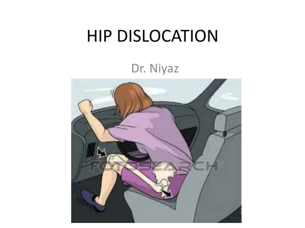
5. AETIOLOGY Extreme mouth opening- dental & ENT TX, under GA,yawning
Trauma- falls, RTA
TMJ dx � osteoarthrosis, int.joint derangement
Hypermobility assoc. with systemic dx e.g Ehler�s Danlos syndrome, CT disorders
Malocclusion- Angles class 2 div 1
6. AETIOLOGY Occlusal disharmony- long term over-closure foll. Edentulism, cos of alveolar bone resorption
Ill-fitting denture
Psychogenic & neurological disorders e.g Parkinson�s dx, multiple sclerosis,tardive dyskinesia
Neuroleptic drugs e.g phenothiazine; antiemetic e.g metoclopromide (extrapyramidal effects? spasms of jaws & facial muscles
Px with congenitally shallow GF or underdeveloped condyle
7. DIAGNOSIS History of factors causing occlusal disharmony, use of neuroleptic drugs, presence of psychogenic or neurological problems, hyperextension of other joints, familial hx of dislocation
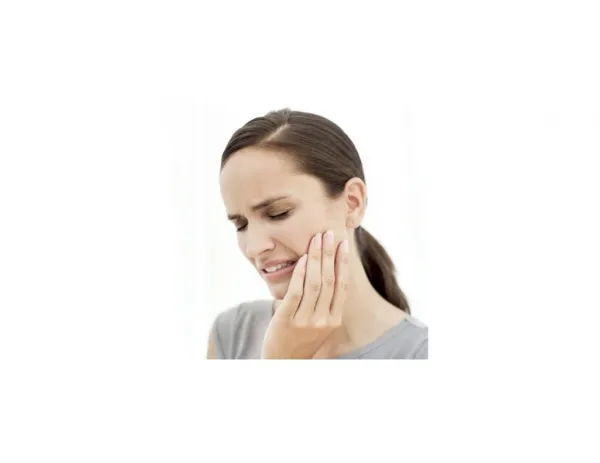
Examination � check for mandibular prognathism, hollow ant to tragus, palpable CH ant to AE, ant open bite, limited mouth opening, pain in or around TMJ
8. CLASSIFICATION Various exists
Acute, chronic & recurrent (Rowe& Killey 1968)
Anterior (Heslop 1956)- CH moves ant to AE; antero- lat variant (moris & Hutton (1957)
Posterior (Helmy 1957)- movt of CH posteriorly, assoc base of skull # or ant wall of bony meatus
9. CLASSIFICATION Lateral (Attery & Young 1969)-2 types:
Type1 �lateral subluxation
Type2 � complete dislocation with CH forced laterally & superiorly into temporal fossa ( assoc with parasymphyseal #)
Superior (Zeccha 1977)- displacement of CH into middle cranial fossa ( assoc with # of GF)
10. INVESTIGATION Plain radiographs (TMJ views)� transcraniooblique; reverse towne�s; others: PA ,R & L oblique laterals
Conventional tomograms� orthopantomogram; plain tomograms (lat)
Computerized tomograms-3D CT scans
MRI
Ultrasound
11. TREATMENT-nonsurgical & surgical methods Non-surgical methods
Bimanual reduction with or without anxiolytics &/or LA, GA. Rest jaw 2-3wks
Slow elastic traction with Erich pattern arch bars & post bite plane. Rest jaw for 2-3 weeks
Chemical capsulorrhaphy using Na psylliate (Schultz 1949) 0.5% 1ml soln of Na tetradecyl sulphate(STD) 3X 2-6wk interval ? causes pericapsular fibrosis which limit CH excursion
Injection of autologous blood into the joint Injection of botulinium toxin type A
12. SURGICAL METHODS Restitution of ligaments & plication of capsule (surgical capsulorrhaphy). Suture line reinforced by turning down a flap of temporal fascia and securing this to both capsule & ligament
Limitation of forward movt by ligation of condyle- tying a length of fascia lata or mersilene (Georgiade 1965) both to zygomatic arch & around condylar neck
13. SURGICAL METHODS Limitation of forward movt by augmentation of AE using:
bone graft from zygomatic arch, mastoid process, iliac crest & calvarium
Calvarial graft- high quality, low incidence of resorption, minimal donor site morbidity, etc, contain large amount of bone
Allele graft- L-shaped SS pins (Findlay 1964); vitallium mesh (Howe& Kent 1978); titallium miniplates (silastic implants
14. SURGICAL METHODS Down fracturing of zygomatic arch
Mayer (1933)
Leclerc & Girard (1943) vertical osteotomy
Dautrey & Gosserez (1964) post. slanting osteotomy
Elimination of dislocation by removal of AE (Myrrhaug (1951)- px continues to dislocate but reduction by px is automatic & painless-condylectomy(Reidel, condylotomy
15. SURGICAL METHODS Prevention of dislocation by removal of activating muscle
Myotomy- lat pterygoid myotomy+/- discectomy
Temporalis myotomy (Laskin)
16. COMPLICATIONS Relapse / recurrence
Facial nerve palsy
Limited mouth opening
Infection
Pseudoarthrosis
Scar formation