Download
1 / 13
140 likes | 276 Views
PERIPHERAL OSSIFYING FIBROMA. OSSIFYING FIBROID EPULIS. PERIPHERAL FIBROMA WITH CALCIFICATION. CALCIFYING FIBROBLASTIC GRANULOMA. a relatively common gingival growth reactive rather than neoplastic in nature. uncertain pathogenesis. clinical and histopathologicSimilarities

E N D
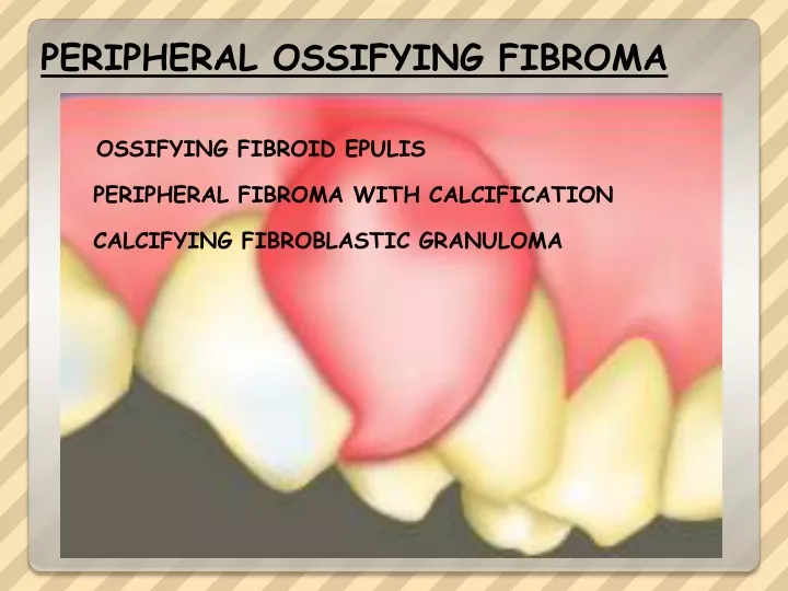
PERIPHERAL OSSIFYING FIBROMA OSSIFYING FIBROID EPULIS PERIPHERAL FIBROMA WITH CALCIFICATION CALCIFYING FIBROBLASTIC GRANULOMA
a relatively common gingival growth reactive rather than neoplastic in nature uncertain pathogenesis clinical and histopathologicSimilarities some POF are………fibrous maturation and subsequent calcification of a pyogenic granulomas However, not all POFs may develop in this manner origin of the mineralized product……. Probably from cells of the periosteum or periodontal ligament. does not represent the soft tissue counter-part of the central ossifying fibroma
occurs exclusively on the gingiva a nodular mass, Pedunculated or sessile usually from the interdental papilla red to pink color frequently ulceratedRed ulcerated lesions often are mistaken for pyogenic granulomas Most lesions <2 cm Pyogenic granuloma
These are usually associated with local irritation such as calculus or mal-alignment of teeth. a somewhat nodular, firm lesion covered by intact squamous epithelium. little excessive vascular proliferation
Teenagers and young adults Almost two thirds of all cases occur in females a slight predilection for the maxillary arch Rarely migration and loosening of adjacent teeth A large lesion
Histopathologic Features a fibrous proliferation associated with the formation of a mineralized product If the epithelium is ulcerated, the surface is covered by a fibrinopurulent membrane with a subjacent zone of granulation tissue.
The deeper fibroblastic component often is cellular , especially in areas of mineralization
formation of bone within a moderately cellular fibrous stroma.
mineralized component …………… bone, cementum-like material, or dystrophic calcifications . In some cases, the fibroblastic proliferation and associated mineralization is only a small component of a larger mass that resembles a fibroma or pyogenic granuloma. immature metaplastic bone.
Usually, the bone is woven and trabecular in type, although older lesions may demonstrate mature lamellar bone.
dystrophic calcifications multiple granules, tiny globules, or large, irregular masses of basophilic mineralized material. dystrophic calcifications are more common in early, ulcerated lesions