Download
1 / 23
230 likes | 411 Views
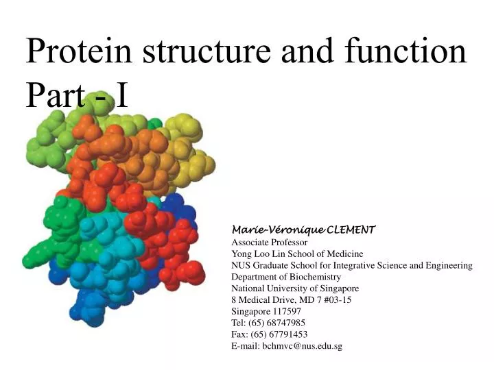
Protein structure and function Part - I. Marie-Véronique CLEMENT Associate Professor Yong Loo Lin School of Medicine NUS Graduate School for Integrative Science and Engineering Department of Biochemistry National University of Singapore 8 Medical Drive, MD 7 #03-15 Singapore 117597

E N D
Protein structure and function Part - I Marie-Véronique CLEMENT Associate Professor Yong Loo Lin School of Medicine NUS Graduate School for Integrative Science and Engineering Department of Biochemistry National University of Singapore 8 Medical Drive, MD 7 #03-15 Singapore 117597 Tel: (65) 68747985 Fax: (65) 67791453 E-mail: bchmvc@nus.edu.sg
N-terminus terminates by an amino group Peptide bond Amino acid C-terminus terminates by a carboxyl group From amino acids to protein: A peptide: Phe-Ser-Glu-Lys (F-S-E-K)
The Shape of proteins: Occurs Spontaneously Native conformation determined by different Levels of structure
Four Levels of Structure Determine the Shape of Proteins Primary structure The linear arrangement (sequence) of amino acids and the location of covalent (mostly disulfide) bonds within a polypeptide chain. Determined by the genetic code. Secondary structure local folding of a polypeptide chain into regular structures including the a helix, b sheet, and U-shaped turns and loops. Tertiary structure overall three-dimensional form of a polypeptide chain, which is stabilized by multiple non-covalent interactions between side chains. Quaternary structure: The number and relative positions of the polypeptide chains in multisubunit proteins. Not all protein have a quaternary structure.
Primary Structure of a protein: determined by the nucleotide sequence of its gene Bovine Insulin: the first sequenced protein • In 1953, Frederick Sanger determined the amino acid sequence of insulin, a protein hormone . • This work is a landmark in biochemistry because it showed for the first time that a protein has a precisely defined amino acid sequence. • it demonstrated that insulin consists only of amino acids linked by peptide bonds between α-amino and α-carboxyl groups. • the complete amino acid sequences of more than 100,000 proteins are now known. • Each protein has a unique, precisely defined amino acid sequence.
Primary Structure Pro-insulin is produced in the Pancreatic islet cells C-peptide Pro-insulin protein 30/31 65/66 Human: Thr-Ser-Ile Cow: Ala-Ser-Val Pig: Thr-Ser-Ile Chiken: His-Asn-Thr Insuline + C peptide C-peptide
Amino acid substitution in proteins from different species Substitution of an amino acid by another amino acid of similar polarity (Val for Ile in position 10 of insulin) Conservative Substitution involving replacement of an amino acid by another of different polarity (sickle cell anemia, 6th position of hemoglobin replace from a glutamic acid to a valine induce precipitation of hemoglobin in red blood cells) Non conservative Invariant residues Amino acid found at the same position in different species (critical for for the sructure or function of the protein)
Protein conformation: most of the proteins fold into only one stable conformation or native conformation More than 50 amino acids becomes a protein
SECONDARY STRUCTURE • Stabilized by hydrogen bonds • H- bonds are between –CO and –NH groups of peptide backbone • H-bonds are either intra- or inter- molecular • 3 types : a-helix, b-sheet and triple-helix
What forces determine the structure? • Primary structure - determined by covalent bonds • Secondary, Tertiary, Quaternary structures - all determined by weak forces • Weak forces - H-bonds, ionic interactions, van der Waals interactions, hydrophobic interactions
Secondary structures: a Helix: a helix conformation was discovered 50 years ago in a keratine abundant in hair nails, and horns b Sheet: discovered within a year of the discovery of a helix.Found in protein fibroin the major constituant of silk
The a helix: result from hydrogen bonding, does not involve the side chain of the amino acid
sheet: result from hydrogen bonding, does not involve the side chain of the amino acid
Two type of b Sheet structures An anti paralellel b sheet A paralellel b sheet
TRIPLE HELIX • Limited to tropocollagen molecule • Sequence motif of –(Gly-X-Pro/Hypro)n- • 3 left-handed helices wound together to give a right-handed superhelix • Stable superhelix : glycines located on the central axis (small R group) of triple helix • One interchain H-bond for each triplet of aas – between NH of Gly and CO of X (or Proline) in the adjacent chain
NONREPETITIVE STRUCTURES • Helices/b-sheets: ~50% of regular 2ostructures of globular proteins • Remaining : coil or loop conformation • Also quite regular, but difficult to describe • Examples : reverse turns, b-bends (connect successive strands of antiparallel b-sheets)
The Beta Turn (aka beta bend, tight turn) • allows the peptide chain to reverse direction • carbonyl O of one residue is H-bonded to the amide proton of a residue three residues away • proline and glycine are prevalent in beta turns (?)
b-bulge • A strand of polypeptide in a b-sheet may contain an “extra” residue • This extra residue is not hydrogen bonded to a neighbouring strand • This is known as a b-bulge.
Tertiary structure: the overall shape of a protein or a telephone cord!!! The secondary structure of a telephone cord A telephone cord, specifically the coil of a telephone cord, can be used as an analogy to the alpha helix secondary structure of a protein. The tertiary structure of a telephone cord The tertiary structure of a protein refers to the way the secondary structure folds back upon itself or twists around to form a three-dimensional structure. The secondary coil structure is still there, but the tertiary tangle has been superimposed on it.
Tertiary structure: the overall shape of a protein Full three dimensional organization of a protein The three-dimensional structure of a protein kinase