Download
1 / 16
160 likes | 252 Views
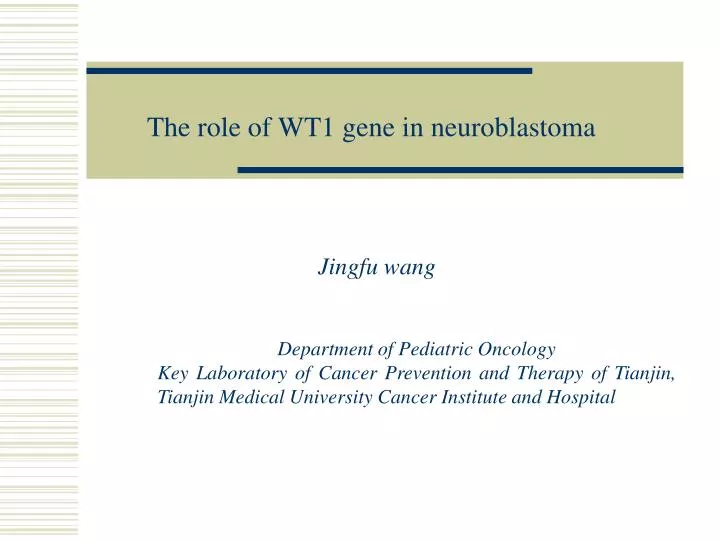
The role of WT1 gene in neuroblastoma. Jingfu wang. Department of Pediatric Oncology Key Laboratory of Cancer Prevention and Therapy of Tianjin, Tianjin Medical University Cancer Institute and Hospital. Background /purpose.

E N D
The role of WT1 gene in neuroblastoma Jingfu wang Department of Pediatric Oncology Key Laboratory of Cancer Prevention and Therapy of Tianjin, Tianjin Medical University Cancer Institute and Hospital
Background/purpose • The recurrence of neuroblastoma due to minimal residual disease (MRD) isoften seen.Immunotherapy is an attractive therapeutic option forcontrolling MRD. • Wilms tumor gene (WT1) was firstly identified as a suppressor involved in the development of Wilms tumor. However, oncogenic properties of WT1 were recently observed in various hematological and solid malignancies. • Over the past years, the immunotherapy using peptide vaccines against WT1 in adults with leukemia and various solid cancers was promising. • To determine whether WT1 vaccines are applicable for neuroblastoma, firstly, we must understand the exact function of WT1 in neuroblastoma. So the aim of this study was to probe it.
Materials and methods • WT1 mRNA expression in the tumor tissue of 22 neuroblastomas (NBs), 5 ganglioneuromas (GNs) and 4 NB cell lines:conventional and real-time RT-PCR. • WT1 protein expression in 20 NBs and 5 GNs: immunohistochemistry. • Effect of WT1 gene knockdown on NB cell (NB19 and NB69) proliferation: siRNA against WT1; WST-1 assay.
Materials and methods (continue) F: forward; R: reverse; P: probe.
2.50E-02 2.00E-02 1.50E-02 1.00E-02 5.00E-03 0.00E+00 GN NB WT1 mRNA expression in NBs and GNs Correlation of WT1 mRNA levels and histological grade Comparison between neuroblastoma and ganglioneuroma Mann-Whitney Test p=0.261 There was no difference between neuroblastoma and ganglioneuroma
2.50E-02 2.00E-02 1.50E-02 1.00E-02 5.00E-03 0.00E+00 Ⅰ Ⅱ Ⅲ Ⅳ WT1 mRNA expression in NBs and GNs (continue) Correlation of WT1 mRNA levels and clinical stage Comparison among Stage Ⅰ,Ⅱ, Ⅲ and Ⅳ in neuroblastoma Kruskal-Wallis Test P=0.412 There was no difference among stage groups
WT1 mRNA expression in NBs and GNs (continue) Correlation of WT1 mRNA levels and prognosis Log-Rank test:p=0.193 The WT1 mRNA expression levels did not affect the prognosis Median: 8.55×10⁻⁴
WT1 mRNA expression in neuroblastic cell lines The highest expression appeared in NB69 without MYCN amplification (relatively low malignancy)
Summary1 • The levels of WT1 mRNA expression were not correlated with histological grade, clinical stage and prognosis. • Among the four neuroblastic cells, NB69 with relatively low malignancyexhibited the highest WT1 mRNA expression. WT1 does not play a significant role in the oncogenicity of neuroblastoma.
WT1 protein expression in NBs andGNs Neuroblastic cells Ganglion cells WT1 proteins were more strongly expressed in mature ganglion cells than neuroblastic cells
WT1 protein expression in NBs andGNs(continue) Fisher's Exact Probability Test: *p=0.000; ** p=0.009. NB: neuroblastoma; GN: ganglioneuroma. The positive rate was significantly higher in GNs than in NBs
Summary2 • Higher WT1 protein expression in ganglion cells and higher positive rate in GNs provides a clue that WT1 protein may be a candidate factor inducing primitive neuroblastic cells to differentiate into mature ganglion cell.
Effect of WT1 gene knockdown on NB cell (NB19 and NB69) proliferation A and B, Conventional RT-PCR (A) and real-time RT-PCR (B) revealed that WT1 gene was obviously knocked down in NB19 and NB69 cells. C and D, There was no significant change on cell proliferation of NB19 between negative control and WT1 siRNA group (* p=0.937). However, silencing of WT1 gene prompted cell growth in NB69 cell which possessed the highest WT1 mRNA expression (** p=0.001).
Summary3 • The silence of WT1 gene promoting cell growth in NB69 cells notes that WT1 may be a factor inhibiting neuroblastic cells growth.
In conclusion • WT1 may be related to cell differentiation and suppression of cell proliferation in NB. • WT1 gene does not act as an oncogene, but participates in the maturation of NB.
Epithelial cells Mesenchymal cells How to understand our findings contrast to that in adults A common role of WT1: regulating the mesenchymal-epithelial balance The adult cancers with high WT1 expression are generally derived from epithelial cells. These tumors will undergo an epithelial-mesenchymal transition (EMT), and this is often related to a worse prognosis. In contrast, neuroblastoma is derived from primitive mesenchymal cells and WT1 normally plays a role of mesenchymal-epithelial transition (MET). The effect of forcing the cells towards an epithelial state is linked to a favorable prognosis.