Download
1 / 1
10 likes | 92 Views
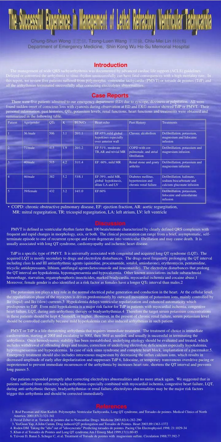
Patient. Age/gender. QTc. K. BUN/Cr. Heart echo. Past History. Treatments. 1. 36 /male. 506. 3.1. 20/1.1. EF:45%,mild global hypokines especially over anterior wall. Chronic alcoholism. Defibrillation, potassium, magnesium and lidocaine infusion. 2. 71/male. 415. 1.9. 20/1.2.

E N D
Patient Age/gender QTc K BUN/Cr Heart echo Past History Treatments 1 36 /male 506 3.1 20/1.1 EF:45%,mild global hypokines especially over anterior wall Chronic alcoholism Defibrillation, potassium, magnesium and lidocaine infusion 2 71/male 415 1.9 20/1.2 EF:51%, moderate TR,AR and trivial MR COPD with cor-pulmonale, and atrial fibrillation Defibrillation, potassium and magnesium infusion. 3 40/male 515 4.2 31/1.4 EF: 60%, mild MR Renal stone and gouty arthritis Defibrillation, potassium and magnesium infusion 4 46/male 382 5.2 53/8.1 EF:39%, mild MR, global hypokinesis, dilate LA and LV Diabetes mellitus, hypertension and chronic renal failure Defibrillation, kalimate, sodium biocarbonate and calcium gluconate infusion 5 39/female 432 3.2 14/1.0 EF:60% - Defibrillation, potassium, lidocaine and amiodarone infusion The Successful Experience in Management of Lethal Refractory Ventricular Tachycardia Chung-Shun Wong 王忠信, Tzong-Luen Wang 王宗倫, Chiu-Mei Lin 林秋梅 Department of Emergency Medicine, Shin Kong Wu Ho-Su Memorial Hospital Introduction The management of wide QRS tachyarrhythmia was documented by advanced cardiac life support (ACLS) guidelines. Delayed or converted the arrhythmia to sinus rhythm unsuccessfully can have fatal consequences with a high mortality rate. In this report, we review five patients suffered from polymorphic ventricular tachycardia (PMVT) or torsade de pointes (TdP) and all the arrhythmias terminated successfully after correcting electrolytes abnormalities. Case Reports There were five patients admitted to our emergency department (ED) due to syncope, dizziness or palpitation. All were found sudden onset of conscious loss with cyanosis during observation at ED and EKG monitor showed TdP or PMVT. Their personal information, past histories, QTc, potassium levels, renal functions, heart functions and treatments were obtained and summarized in the following table. • COPD: chronic obstructive pulmonary disease, EF: ejection fraction, AR: aortic regurgitation, • MR: mitral regurgitation, TR: tricuspid regurgitation, LA: left atrium, LV: left ventricle Discussion PMVT is defined as ventricular rhythm faster than 100 beats/minute characterized by clearly defined QRS complexes with frequent and rapid changes in morphology, axis, or both. The clinical presentation can range from a brief, asymptomatic, self-terminate episode to one of recurrent syncope and even degenerate into ventricular fibrillation and may cause death. It is usually associated with long QT syndrome, cardiomyopathy and ischemic heart disease. TdP is a specific type of PMVT. It is universally associated with congenital and acquired long QT syndrome (LQT). The acquired LQT is mostly secondary to drugs and electrolyte disturbances. The drugs most frequently prolonging the QT interval including class Ia and III antiarrhythmic agents (quinidine, procainamide, sotalol, amiodarone), erythromycin, pentamidine, tricyclic antidepressants, lithium, antifungal agents(ketoconazole and itraconazole). The electrolyte disturbances that prolong the QT interval are hypokalemia, hypomagnesaemia and hypocalcemia. Other known associations include subarachnoid hemorrhage, starvation, organophosphoris insecticides, marked bradycardia, myocardial ischemia and cocaine abuse.1 Moreover, female gender is also identified as a risk factor as females have a longer QTc interval than males.2 The potassium ion plays a key role in the normal electrical pulse generation and conduction in the heart. At the cellular level, the repolarisation phase of the myocytes is driven predominantly by outward movement of potassium ions, mainly controlled by Ikr (rapid) and Iks (slow) currents.3 Hypokalemia delays ventricular repolarization and enhanced automaticity which predisposes to TdP. Even mild hypokalemia is a major risk factor for TdP in patients with myocardial ischemia, congestive heart failure, LQT, during anti-arrhythmic therapy or bradyarrhythmias.4 Therefore the target serum potassium concentrations in these patients should be kept 4.5mmol/L or higher. However, in the present of chronic renal failure, serum potassium level should be controlled carefully because mild hyperkalemia can also induced arrhythmias in such patient. PMVT or TdP is a life-threatening arrhythmia that requires immediate treatment. The treatment of choice is immediate cardioversion, starting at 200J and escalating to 300J, then 360J as needed, and usually is successful in terminating the arrhythmia. Once hemodynamic stability has been reestablished, underlying etiology should be evaluated and treated, which includes withdrawal of offending drugs and toxins, correction of underlying electrolyte deficiencies especially hypokalemia, hypomagnesaemia and hypocalcemia. In case of sick sinus syndrome and complete heart block, implantation of a pacemaker.5. Emergency treatment should also includes intravenous magnesium by decreasing the influx calcium ions, which results in decreased amplitude of early after depolarization and suppresses TdP 6, lidocaine, or temporary transvenous overdrive pacing or isoproterenol to prevent immediate recurrences of the arrhythmia by increases heart rate, shortens the QT interval and prevents long pauses 5. Our patients responded promptly after correcting electrolytes abnormalities and no more attack again. We suggested that in patients suffered from refractory tachyarrhythmia especially combined with myocardial ischemia, congestive heart failure, LQT, during anti-arrhythmic therapy, bradyarrhythmias or renal failure, electrolytes abnormalities may be the major risk factors trigger this arrhythmia and should be corrected immediately. References 1. Rod Passman and Alan Kadish. Polymorphic Ventricular Tachycardia, Long QT syndrome, and Torsades de pointes. Medical Clinics of North America. 2001;85(2):321-341 2. David Zelter et at. Torsade de pointes due to Noncardiac Drugs. Medicine 2003;82(4):282-290 3. YeeGuan Yap, A John Camm. Drug induced QT prolongation and Torsades de Pointes. Heart 2003;89:1363-1372 4. Roden DM. Taking the “idio” out of “idiosyncratic” Predicting torsades de pointes. Pacing Clin Electrophysiol.1998; 21:1029-34 5..Khan et al.Torsades de Pointes and Long QT syndrome.Am J Emerg Med 1999;17(1):80-85 6. Tzivoni D, Banai S, Schuger C, et al. Treatment of Torsade de pointes with magnesium sulfate. Circulation 1988;77:392-7