Download
1 / 64
690 likes | 737 Views
Light and Sight. How do we see?. Electromagnetic Waves: Seeing Objects and Color. Essential Question: How are wavelengths detected by the human eye?. Sight. Visible light is the part of the electromagnetic spectrum that you can see. Remember………………. The Electromagnetic Spectrum.

E N D
Electromagnetic Waves: Seeing Objects and Color Essential Question: How are wavelengths detected by the human eye?
Sight • Visible light is the part of the electromagnetic spectrum that you can see. • Remember………………
The Electromagnetic Spectrum The electromagnetic spectrum is the range of electromagnetic waves extending from radio waves to gamma rays Increasing frequency R O Y G B I V
The colors of visible light are created by electromagnetic energy of various wavelengths (frequencies). See below White light is made up of all the wavelengths of visible light.
Electromagnetic Waves: Seeing Color When white light is refracted, it can be separated into its component colors. As light passes through a prism, refraction causes light to bend and separate into many colors.
A rainbow is produced when a raindrop acts like a prism causing white light to refract (bend) and separate into many colors
Electromagnetic Waves:Seeing Color • Humans see different wavelengths of light as different colors. • Humans see long wavelengths as red • Humans see short wavelengths as violet • Some colors, like pink and brown, are seen when certain combinations of wavelengths are present.
Electromagnetic Waves:Seeing Color If we see an object because light is reflected off the object by a light source and white light is made up of all the wavelengths of visible light, why do objects have different colors? The color of an object is determined by the wavelengths (color) of light it reflects. So, if an object reflects one wavelength (color), it absorbs all the other wavelengths (colors) of visible light.
In this example, the sun is the light source. The sun’s light appears white because it is made up of all the wavelengths of visible light. However, humans see the apple as red because all of the other wavelengths (or colors) are absorbed by the apple. The wavelength that we see as red is reflected off the apple.
Seeing Color • An object that reflects all colors makes it look white • An object that reflects none of the light and only absorbs it looks black
We only see a very small portion of the sun’s energy. • So, what is the path that this energy travels so that we can see?
BINOCULAR VISION • Humans have binocular vision…vision where both eyes aim at the same visual target, at the same time; both eyes work together -- simultaneously, equally and accurately -- as a coordinated team.
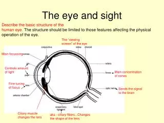
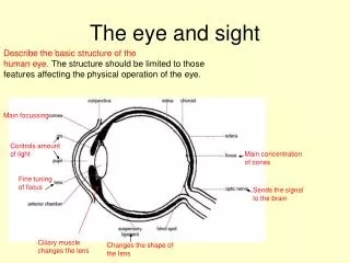
Pupil Lens Iris Cornea Retina Sclera Conjunctiva Ciliary body Vitreous body Aqueous chamber Optic nerve Rods Cones Blind spot Parts of the Eye
Sclera and Cornea • The sclera, or white of the eye, forms the outermost layer of the eyeball. • The sclera consists of a firm fibrous membrane that maintains the shape of the eye and gives attachment to the extrinsic musclesof the eye.
Anteriorly the sclera continues as a clear transparent epithelial membrane, the cornea. • Light rays pass through the cornea to reach the retina. • The cornea is involved in refracting (bending) light rays to focus them on the retina.
Ciliary Body • The ciliary body consists of ciliary muscle (smooth muscle fibers) and secretory epithelial cells. • It gives attachment to the suspensory ligament which, at its other end, is attached to the capsule enclosing the lens. • Contraction and relaxation of the ciliary muscle changes the thickness of the lens, which bends light rays entering the eye to focus them on the retina.
Iris • The iris is the visible colored part of the eye and lies behind the cornea and in front of the lens. • It divides the anterior segment of the eye into anterior and posterior chambers which contain the fluids of the eye.. • It is a circular body composed of pigment cells and two layers of smooth muscle fibers, one circular and the other radiating. • In the center of the iris is called the pupil.
The iris is supplied by nerves. • Stimulation of the nerves can constrict or dilate the pupil . • The color of the iris is genetically determined and depends on the number of pigment cells present. • Albinos have no pigment cells and people with blue eyes have fewer than those with brown eyes.
Lens • The lens is a highly elastic circular biconvex body, lying immediately behind the pupil. • It consists of fibers enclosed within a capsule and it is suspended from the ciliary body by the suspensory ligament. • Its thickness is controlled by the ciliary muscle through the suspensory ligament.
When the ciliary muscle contracts, it moves forward, releasing its pull on the lens, increasing its thickness. • The nearer is the object being viewed, the thicker the lens becomes to allow focusing. • The lens refracts light rays reflected by objects in front of the eye.
Retina • The retina is the innermost layer of the wall of the eye. • It is an extremely delicate structure and is well adapted for stimulation by light rays. • It is composed of several layers of nerve cell bodies
Near the center of the posterior part is the macula lutea, or yellow spot. • In the centre of the yellow spot is a little depression called the fovea centralis, consisting of only cones. This is where vision is greatest.
The light-sensitive layer consists of sensory receptor cells: rods and cones. • The rods and cones contain photosensitive pigments that convert light rays into nerve impulses. • Rods see white and black. • Cones see color. There are 3 colored cones: Red, Blue, and Green.
The small area of retina where the optic nerve leaves the eye is the optic disc or blind spot. • It has no light sensitive cells
Interior of the eye • The space between the cornea and the lens, is incompletely divided into anterior and posterior chambers by the iris. • Both chambers contain a clear aqueous fluid secreted into the posterior chamber by ciliary glands.
There is continuous production and drainage but the intraocular pressure remains fairly constant An increase in this pressure causes glaucoma. • Aqueous fluid supplies nutrients and removes wastes from the transparent structures in the front of the eye that have no blood supply, i.e. the cornea, lens and lens capsule.
Behind the lens of the eyeball is the vitreous body. • This is a soft, colorless, transparent, jelly-like substance composed of 99% water, some salts and mucoprotein. • The vitreous body maintains intraocular pressure to support the retina and prevent the eyeball from collapsing or changing shape.
Yellow Spot(Fovea Centralis) Scleratic Coat(Sclera) Retina Iris Macula Cornea Choroid Coat(Choroid Membrane) Optic Nerve Pupil Vitreous Lens Ciliary Muscle DIAGRAM OF THE EYE
Ciliary Body Blind spot
Lens Introduction • Lenses are very useful. e.g. in cameras, projectors, telescopes, microscopes and eyes • There are two types of lenses that we will talk about : -convex -concave
CONCAVE or NEGATIVE lenses will DIVERGE (spread out) light rays DRAW IT!
Concave lens The correct name of nearsightedness is myopia.Myopia occurs when the eyeball is slightly longer than usual from front to back. This causes light rays to focus at a point in front of the retina, rather than directly on its surface. A concave lens is usually used to correct this problem.
Convexlens The correct name for farsightedness is Hyperopia. The shape of your eye does not bend light correctly, resulting in a blurred image. A convex lens is usually used to correct this problem.