Download
1 / 88
880 likes | 892 Views
Toxicology course. Part 2: General approaches to the management poisoned patients. Poisonings and drug overdoses can cause quick physical and mental changes in a person. Bystanders usually are the ones who must initiate care and call a poison control center or emergency number.

E N D
Toxicology course Part 2: General approaches to the management poisoned patients N.B
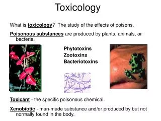
Poisonings and drug overdoses can cause quick physical and mental changes in a person. • Bystanders usually are the ones who must initiate care and call a poison control center or emergency number. • Chemical agents that cause toxicity include: • Drugs • Insecticides/herbicides • Plant toxins, Animal toxins • Chemical weapons, • Radioactive elements N.B
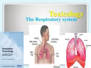
Most exposures to toxic fumes occur in the home.Burning wood, gas, oil, coal, or kerosene produces carbon monoxide (CO). • CO gas is colorless, odorless, tasteless, and non-irritating, which makes it especially dangerous. • Cellular hypoxia may occur in spite of adequate ventilation and oxygen administration when poising is due to carbon monoxide, cyanide, hydrogen sulfide, and other poisons that interfere with transport or utilization of oxygen.
In such patients, cellular hypoxia is evident by the development of tachycardia, hypotension, severe lactic acidosis, and ischemia. • Commonly observed poisonings or drug overdoses are caused by (but certainly not limited to) carbon monoxide, salicylates, acetaminophen, nicotine, alcohol, heroin, marijuana, narcotic analgesics, benzodiazepines, tricyclic antidepressants, amphetamines, and cocaine.
Approach to the poisened patient • How does the poisoned patient die? • Many toxins depress the Central Nervous System(CNS), resulting in coma. • Patients under the influence of hallucinogens such as LSD may die in fights or falls from high places.
Comatose patients frequently lose their airway protective reflexes and their respiratory drive. Thus they may die as a result of airway obstruction by the flaccid tongue, aspiration of gastric contents into the tracheobronchial tree, or respiratory arrest . • These are the most common causes of death due to overdose of narcotics and sedative-hypnotic drugs.
Cardiovascular toxicity is also frequently encountered in poising. Hypotension may be due to depression of cardiac contractility; peripheral vascular collapse due to blockade of alpha adrenoceptor-mediated vascular tone or cardiac arrhythmias. • Lethal arrhythmias such as ventricular tachycardia and fibrillation can occur with overdoses of many cardioactive drugs such as epinephrine, amphetamines, cocaine, digitalis and theophylline; and drugs not usually considered cardioactive, such as tricyclic antidepressants, antihistamines, and some opioid analogs.
Hypothermia or hyperthermia due to exposure as well as the temperature dysregulating effects of many drugs can also produce hypotension. • Hyperthermia may result from sustained muscular hyperreactivity and can lead to muscle breakdown and myoglobinuria, renal failure, lactic acidosis, and hyperkalemia.
Seizures, muscular hyperactivity, and rigidity may result in death. • Seizures may cause pulmonary aspiration,hypoxia, and brain damage. • Drugs and poisons that often cause seizures include antidepressants, isoniazid, diphenhydramine, cocaine, and amphetamines.
Some organ system damage may occur after poisoning and is sometimes delayed in onset. • Pulmonary fibrosis may begin several days after ingestion. • Massive hepatic necrosis due to poisoning by acetaminophen or certain mushrooms result in hepatic encephalopathy and death 48-72 hours or longer ingestion.
CLINICAL STRATEGY FOR THETREATMENT OF THE POISONED PATIENT • important elements of the initial clinical encounter for a poisoned patient include: 1. Clinical stabilization of the patient 2. Clinical evaluation (history, physical, laboratory, radiology) 3. Prevention of further toxicant absorption 4. Enhancement of toxicant elimination 5. Administration of antidote (if available) 6. Supportive care, close monitoring, and clinical follow-up N.B
Clinical Stabilization (Resuscitation) • Assessment of ABC’s: • The patient’s Airway(ability to move air into and out of the lungs) should be cleared of any obstruction, oral airway or nasotracheal or endotracheal intubation may be necessary to adequately maintain and protect the patient’s airway. • For many patients is sufficient to move the tongue out of the airway. N.B
Breathing (the presence of spontaneous respirations),measuring arterial blood gases, Mechanical ventilation may be necessary to support the patient (ex: heroin depress the respiratory system) • Circulation(adequate blood pressure and perfusion of vital organs) is the initial step of emergency treatment, monitoring of pulse rate, and urinary output • In these cases, fluid balance needs to be carefully controlled. N.B
Invasive monitoring (e.g., central venous pressure, pulmonary artery catheter, Foley catheter with urometer) and drug therapy may be necessary to prevent or minimize complications such as pulmonary edema 2. Vital signs and hypoglycemia • Giving dextrose to correct hypoglycemia N.B
Most poisoned patients, with a toxic exposure, will exhibit symptoms early in their presentation….. Some don’t show significant symptoms for a period of time. • Ex: Some drugs, such as a benzodiazepine, can cause significant sedation early after exposure but often have a comparatively mild clinical course, whereas camphor, show little clinical effects initially but can produce a fatal outcome. N.B
Clinical evaluation (history, physical, laboratory, radiology) • History: • Determine the substance, dose, route • The extent of exposure • Timing of exposure. (most difficult) • Difficulty in obtaining history appeared if the patient is unresponsive and unable to provide the history, suicide or patient who has taken illegal substances is not willing to provide an accurate history. N.B
informative clinical history can be obtained by: • interviewing with family members • emergency medical technicians who were at the scene • a pharmacist who can sometimes provide a listing of prescriptions recently filled • An employer who can provide a list of chemicals found in the work environment for an occupational exposure. • Check for empty bottles or containers, smells or unusual containers, or suicide not N.B
Physical Examination • Check clothing for objects or substances • Assess general appearance of pt • Agitation, confusion…… • Exam skin for bruising, cyanosis, flushing • Ex: Excessive sweeting occurs with organophosphates, nicotine and sympathomimetic drugs N.B
Exam eyes for pupils size, reactivity, increased lacramaiton….. • Miosis is typical of opioids, cholinesterase inhibitors (e.g. Organophosphate insecticides), and deep coma due to sedative drugs. • Mydriasis is common with amphetamines, cocaine, atropine and other anticholinergic drugs. • Horizontal nystagmus is characteristic of intoxication with alcohol and other sedative drugs N.B
Vital signs: Bp, HR,RR, Temperature • Hyperthermia may be associated with sympathomimetics, anticholinergic, salicylates, and drugs producing seizures. • Hypothermia can be caused by any CNS depressant drug. • Cyanosis may be caused by hypoxemia or by methemoglobinemia. • Oropharynx : increase salivation or excessive dryness, Typical odors of alcohol or ammonia , • CV: rhythm, and regularity • Ex: Hypertension and tachycardia are typical with amphetamines, cocain and anticholinergic drugs. • Hypotension and bradycardia are characteristic features of overdose with calcium channel blockers, beta blockers, clonidine, and sedative hipnotics. • Hypotension with tachycardia is common with tricyclic antidepressants, vasodilators and beta agonists. N.B
Lungs: rate,wheezing….. • Ex:Rapid respirations are typical of salicylates, CO poisoning • Abd: bowel sounds, tenderness or rigidity… • Hyperactive bowel sounds, tenderness abdominal cramping and diarrhea are common in poisoning with organophosphatase, iron, arsenic • Ext: tremor….. • Neuro: reflexes, muscle tone coordination…. • Physiologic excitation – anticholinergic, sympathomimetic, drug withdrawal • Physiologic depression –cholinergic (parasympathomimetic), opiate, or sedative-hypnotic agents, or alcohols • muscular rigidity can be caused by anti-psychotic agents, serotonin syndrome N.B
Many factors can affect the patient’s mental status. • Hypoglycemia and hypoxemia; that can be life-threatening but easily addressed by administering oxygen and IV dextrose until laboratory results are available. N.B
A toxidrome is a group of signs and symptoms associated with overdose or exposure to a particular category of drugs and toxins • The major toxic syndromes include narcotic, cholinergic, sympathomimetic, and anticholinergic. • For example, if a patient presents with altered mental status, mydriasis (dilated pupils), mild hypertension, tachycardia, warm skin, dry mucous membranes, and diminished bowel sounds in the abdomen, the clinical toxicologist can characterize the patient’s presentation as consistent with the anticholinergic toxic syndrome N.B
Characteristic odor can be detected on the poisoned patient’s breath or clothing N.B
Laboratory Evaluation • Tests that provide clues to the agent(s) taken by the patient include • arterial blood gases (ABGs) • Electrolytes • Serum osmolality tests • Urinalysis • complete blood count • Electrocardiography • Toxicology screens N.B
1.Arterial Blood Gases (ABGs) • Hypoventilation will result in an elevated PCO2 (hypercapnia) and a low PO2(hypoxia). • The PO2 may also be low with aspiration pneumonia or drug induced pulmonary edema. • Signs of inadequate ventilation or oxygenation includecyanosis, tachycardia, hypoventilation, intercostal muscle retractions, and altered mental status. N.B
2. Electrolytes • Acute poisoning can cause an imbalance in a patient’s electrolyte levels, including sodium, potassium, chloride, bicarbonate, carbon dioxide, magnesium, and calcium. • Serum level measurements are also available for carbamazepine, iron, ethanol, lithium, aspirin, and valproic acid and may be obtained if these agents are suspected in an overdose N.B
In 1975, Rumack and Mathews published a nomogram for acetaminophen poisoning. • This nomogram predicts clinical outcome and is also valuable to guide the clinician in the decision as to whether or not to administer N -acetylcysteine (NAC), an antidote for significant acetaminophen ingestion. • several other drugs including lithium, salicylates, digoxin, iron, phenobarbital, and theophylline. N.B
Calculated clinical parameters: • The anion gap (AG) calculations • The osmol gap calculations. • the patient’s condition is consistent with exposure to chemicals known to cause elevations of these parameters (ie, metabolic acidosis, altered mental status, etc). N.B
Elevated AG; metabolic acidosis (low blood pH and low serum HCO 3 ) N.B
The osmolal gap is the difference between the measured osmolality and the calculated osmolality An elevated osmol gap in the setting of a poisoned patient suggests the presence of an osmotically active substance in the plasma that is not accounted for by the Na, glucose, or BUN concentrations N.B
3. Electrocardiography: • Provideevidence of drugs causing arrhythmias or conductiondelays (e.g., tricyclic antidepressants). 4. Renal function tests • Some toxines have direct nephrotoxic, in other cases renal failure is due to shock. • BUN and creatinine levels should be measured and urinanalysis performed. N.B
5. Radiographic Examination • is relatively limited. This is due primarily to the lack of radiopacity of many oral forms of medication. • Chest radiographs: drug-induced noncardiac pulmonary edema is associated with serious intoxication with salicylates and opioid agonists • Abdominal radiographs: foreign Bodies, batteries, lead, halogenated hydrocarbon (chloroform), cocaine or heroin containers. • The computed head tomography:carbon monoxide has been associated with lesions of the brain consisting of low-density areas in the cerebral white matter and in the basal ganglia N.B
6. Renal function tests • Some toxins have direct nephrotoxic, in other cases renal failure is due to shock or myoglobinuria. • BUN and creatinine levels should be measured and urinanalysis performed.
7. Toxicology screens • toxicology screen is a laboratory analysis of a body fluid or tissue to identify drugs or toxins. Saliva, spinal fluid, and hair may be analyzed, blood or urine samples are used more frequently.Each screen tests for specific drugs or agents. • The sample must also be properly collected, and there must be a laboratory near enough to obtain results quickly. N.B
The test sample must be collected while the drug or toxin is in the body fluid or tissue used for testing. • For example, cocaine is a rapidly metabolized drug; however, its metabolite, benzoylecgonine, can be detected in the urine for several hours after cocaine use. N.B
Management • Prevent absorption of and further exposure to the agent. • Treatment begins with first aid at the scene and continues in the emergency department and often the intensive care unit (ICU). • Advanced management involves further steps to prevent absorption and enhance elimination of the agent. For instance, antidotes, antivenins(the treatment of venomous bites or stings N.B
General- management: • Provision of supportive care • Prevention of poison absorption • Enhancement of elimination of poison • Administration of antidotes N.B
1. Supportive care • Vital signs, mental status, and pupil size • cardiac monitoring, ECG • Protect airway • Intravenous access • Cervical immobilization if suspect trauma • Rule out hypoglycaemia N.B
2. Prevention of Further Poison Absorption Decontamination N.B
To minimize the total amount of chemical that reaches the systemic circulation. • inhalation route: remove the patient from the environment where the toxin is found and to provide adequate ventilation and oxygenation for the patient. • Further evaluation is needed if the patient experiences respiratory irritation or shortness of breath. N.B
2. Topical route: patient clothing containing the chemical must be removed and properly disposed in airtight wrappings or containers to ensure that the rescuers and healthcare providers are adequately protected from secondary exposure. • Require gentle washing of the skin with water and mild soap taking care not to cause cutaneous abrasions of the skin that may enhance dermal absorption. N.B
Some toxins may require further decontamination. For example, three separate soap and water washings or showers are recommended to decontaminate organophosphate pesticides (e.g., Malathion or Diazinon). • Protective clothing should be worn to reduce the risk for toxicity while handling contaminated clothing or assisting with skin decontamination.
Eyes • Many substances can accidentally splash into the eyes. When this happens, the eyes must be flushed to removethe agent. • Immediate irrigation with lukewarm water or normal saline is recommended. • Continuous flooding of the eyes with a large glass of water or low-pressure shower should be done for 15 minutes.
The patient should blink the eyes open and closed during the irrigation. • If necessary, the pH of the eyes can be tested. If the pH is abnormal, irrigation should continue until the pH normalizes. • An ophthalmologic examination is needed when ocular irritation or visual disturbance persists after irrigation.
4. Oral route: Three general methods involve removing toxin from stomach • induction of emesis with syrup of ipecac, gastric lavage, oral administration of activated charcoal, and whole-bowel irrigation. • Many clinical toxicologists believe that there remains a limited role for the clinical use of syrup of ipecac N.B
The accepted contraindications for use of syrup of ipecac are: • Children less than six months of age • in the ingestion of a caustic agent (acid or alkali) • in a patient with a depressed level of consciousness or gag reflex • When there is a significant risk of aspiration of gastric contents such as for ingestion of a liquid hydrocarbon with high aspiration potential N.B
Dosing: 15 ml for 1-12 years old and 30 ml for adults; may repeat once if no emesis in 12 hr. • 90% vomit within 20 minutes of first dose and 97% vomit with second dose. • The American Academy of Pediatrics no longer recommends the use of emetics (such as syrup of ipecac) for GI decontamination. N.B