Download
1 / 10
100 likes | 186 Views
Microscopes. The invention of the microscope in the 17 th century led to the discovery of the cell. Robert Hooke described cells using this light microscope. Modern Microscopes. Visible light is focused on the specimen with the condenser lens. • Light passing through the

E N D
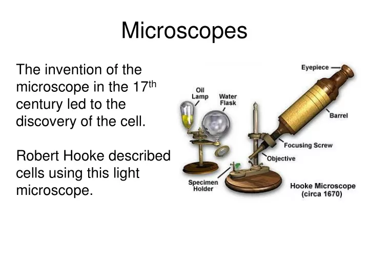
Microscopes The invention of the microscope in the 17th century led to the discovery of the cell. Robert Hooke described cells using this light microscope.
Modern Microscopes • Visible light is focused on the specimen with the condenser lens. • Light passing through the specimen is refracted with an objective and an ocular lens (magnify and invert the image for the observer).
Two Important Properties of Microscopes: • Magnification – how much largeran object is made to appear. • Resolving power – minimum distance between two points that can still be distinguished as two separate points. *limited by the wavelength of light. *maximum resolution of a LM is 0.2μm. *highest magnification is 1,000X.
Gallery of Cells What can you infer about function from the structure of these cells?
Electron Microscopes • Instead of light, electron microscopes use electron beams which have much shorter wavelengths (resolving power of 0.2nm). • Enhanced resolution and magnification led to the identification of subcellular organelles. • There are two types of electron microscopes: transmission and scanning electron microscopes.
Transmission Electron Microscope • Electromagnetic lenses. • Electrons transmitted through the specimen are focused onto a viewing screen or film. • Used to study internal cellular structure.
Scanning Electron Microscope • Electron beam scans the surface of the specimen. • Excites secondary electrons on sample. • Collects secondary electrons and focuses onto a viewing screen. • Great depth of field – 3D image.
http://www.aimediaserver.com/studiodaily/harvard/harvard.swf