Download
1 / 32
880 likes | 3.16k Views
Achalasia. Dan Imler Morning Report. Definition. A Greek term that means "does not relax“ Normally The act of swallowing (deglutition) normally initiates a peristaltic wave that propels ingested material down the esophagus.

E N D
Achalasia Dan Imler Morning Report
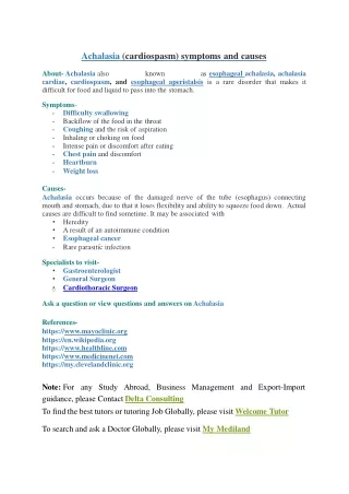
Definition • A Greek term that means "does not relax“ • Normally • The act of swallowing (deglutition) normally initiates a peristaltic wave that propels ingested material down the esophagus. • Deglutition also triggers relaxation of the lower esophageal sphincter (LES), a process that allows the swallowed material to enter the stomach.
Definition • Achalasia is a disease of unknown cause in which there is a loss of peristalsis in the distal esophagus (whose musculature is comprised predominantly of smooth muscle) and a failure of LES relaxation.
Definition • Although both of these abnormalities impair esophageal emptying, the symptoms and signs of achalasia (eg, dysphagia and chest pain) are due primarily to the defect in LES relaxation.
Definition • The relentless LES contraction in achalasia causes functional obstruction of the esophagus that persists until the hydrostatic pressure of the retained material exceeds the pressure generated by the sphincter muscle.
Pathophysiology • Achalasia results from the degeneration of neurons in the esophageal wall. • Histologic examination reveals decreased numbers of neurons (ganglion cells) in the myenteric plexuses, and the ganglion cells that remain often are surrounded by lymphocytes and, less prominently, by eosinophils. • This inflammatory degeneration preferentially involves the nitric oxide-producing, inhibitory neurons that effect the relaxation of esophageal smooth muscle. • However the cholinergic neurons that contribute to LES tone by causing smooth muscle contraction are relatively spared.
Pathophysiology • The disordered motility that characterizes achalasia appears to result primarily from the loss of inhibitory neurons within the wall of the esophagus itself. • Loss of inhibitory innervation in the LES causes the basal sphincter pressure to rise, and renders the sphincter muscle incapable of normal relaxation. • In the smooth muscle portion of the esophageal body, the loss of inhibitory neurons results in aperistalsis.
Cholecystokinin (CCK) test • In normal individuals, the intravenous administration of CCK octapeptide stimulates both the contraction of smooth muscle cells in the LES, and the release of inhibitory neurotransmitters from ganglion cells in the wall of the esophagus. • Thus, the weak, direct stimulatory effect of CCK on the sphincter muscle is opposed by the CCK-induced release of inhibitory neurotransmitters. • The inhibitory effects predominate, and the net result is a fall in LES pressure. • In contrast, when CCK octapeptide is administered to patients with achalasia, the direct stimulatory effect of the hormone on smooth muscle is unopposed and LES pressure rises.
In addition to the LES dysfunction there may also be a subtle defect in reflex relaxation of the upper esophageal sphincter (UES). • The abrupt esophageal distention that results when gas from the stomach suddenly enters the esophagus normally triggers a reflex relaxation of the UES, thereby allowing the gas to escape through the mouth in the form of a belch. • The UES belch reflex can be demonstrated experimentally by injecting air into the esophagus. • In normal subjects, esophageal air injection causes UES relaxation that is accompanied by an audible belch. • In patients with achalasia, however, air injected into the esophagus frequently causes a paradoxical increase in UES pressure without a belch. • This abnormal reflex presumably results from the loss of inhibitory neurons, although the precise neural pathways that effect the reflex are not clear. • The inability to burp in some patients with achalasia may contribute to the esophageal distention and chest pain that often accompany the disease.
Etiology • The cause of the inflammatory degeneration of neurons in achalasia is not known. • The observations that achalasia is associated with HLA-DQw1 and that affected patients often have circulating antibodies to enteric neurons suggest that achalasia may be an autoimmune disorder. • Some investigators have proposed that achalasia may result from chronic infections with herpes zoster or measles viruses, but modern studies have not confirmed an association between achalasia and any recognized viral disease.
Etiology • Malignancy: the most common cause of pseudoachalasia in most populations. In one series, for example, six patients with pseudoachalasia and 161 patients with primary idiopathic achalasia were seen over a 14 year period. • Chagas' disease: seen in Central and South America, esophageal infection with the protozoan parasite Trypanosoma cruzi can result in a loss of intramural ganglion cells leading to aperistalsis and incomplete LES relaxation. • Other causes: A variety of other diseases have been associated with achalasia-like motor abnormalities. These include amyloidosis, sarcoidosis, neurofibromatosis, eosinophilic gastroenteritis, multiple endocrine neoplasia type 2B, juvenile Sjögren's syndrome, chronic idiopathic intestinal pseudo-obstruction, and Fabry disease.
Epidemiology • Achalasia has an annual incidence of approximately 1 case per 100,000. • Men and women are affected with equal frequency. • The disease can occur at virtually any age, but onset before adolescence is decidedly unusual. • Achalasia is usually diagnosed in patients who are between the ages of 25 and 60 years.
Clinical Manifestations • Dysphagia for solids (91 percent) and liquids (85 percent) is the primary clinical feature of achalasia. • Although dysphagia for liquids can occur in patients with other esophageal motility disorders this symptom is most characteristic of achalasia and strongly suggests the diagnosis.
Clinical Manifestations • Difficulty belching is present in approximately 85 percent of patients, although few describe this symptom spontaneously (ie, without specific prompting from the physician). • Weight loss: usually in the range of 5 to 10 kg • Regurgitation of material retained in the flaccid esophagus may occur, especially during recumbency at night, and may result in aspiration. • Chest pain is more common in younger patients, and tends to diminish over the course of several years. • Affected patients may eat more slowly and adopt specific maneuvers such as lifting the neck or throwing the shoulders back in order to enhance esophageal emptying. • Globus sensation (a lump in the throat) has been reported as a presenting symptom. • Hiccups are common and probably result from obstruction of the distal esophagus
Diagnosis • The symptoms of achalasia often are insidious in onset and gradual in progression. • As a result, patients typically experience symptoms for years before seeking medical attention. • In one series of 87 consecutive patients with newly diagnosed achalasia, the mean duration of symptoms was 4.7 years. • The delay in diagnosis was due to misinterpretation of typical findings by physicians rather than atypical clinical manifestations. • Many patients are treated for other disorders such as gastroesophageal reflux disease before the diagnosis of achalasia is made.
Radiographic studies • A barium swallow is the primary screening test when achalasia is suspected on clinical grounds. • The diagnostic accuracy of barium swallow for achalasia is approximately 95 percent. • The barium swallow typically shows a dilated esophagus that terminates in a beak-like narrowing caused by the persistently contracted lower esophageal sphincter (LES). • In some cases, the dilation is so profound that the esophagus assumes a sigmoid shape.
Manometry • Although clinical and radiographic findings may strongly suggest the diagnosis of achalasia, a manometric examination is required for confirmation in virtually all cases. • Elevated resting LES pressure — In the LES, the loss of inhibitory neurons typically causes LES pressures to rise to hypertensive levels. • Incomplete LES relaxation — Normally, there is complete relaxation of the LES after a swallow; in contrast, LES relaxation in response to a swallow may be incomplete or absent in achalasia.
Endoscopy • Endoscopic evaluation is generally recommended for most patients with achalasia primarily to exclude malignancies at the esophagogastric junction that can mimic primary achalasia clinically, radiographically, and manometrically. • Thus recommended for people with symptoms • Duration of symptoms less than six months • Presentation after age 60 • Excessive weight loss in relation to the duration of symptoms • Difficult passage of the endoscope through the gastroesophageal junction
Medical Therapy • No treatment reliably restores function in the body of the esophagus, although the return of peristaltic activity has been observed occasionally after the administration of therapies designed solely to decrease LES pressure. • Nitrates and calcium channel blockers relax the smooth muscle of the LES both in normal individuals and in patients with achalasia, and these agents have been used to treat the disorder with limited success. • The drugs usually are taken sublingually 10 to 30 minutes before meals.
Dilation of the LES • Despite the many variations in technique, most studies describe good to excellent short-term results in 60 to 85 percent of patients with achalasia who are treated with a single session of pneumatic dilation. • Approximately 50 percent of patients with achalasia who are treated initially with a single pneumatic dilation will require further therapy within five years, and that subsequent pneumatic dilations are progressively less likely to result in a sustained remission. • Esophageal perforation is the most common serious complication of pneumatic dilation, occurring in most large series of experienced endoscopists in 2 to 6 percent of cases
Surgical Myotomy • Surgical myotomy via the modified Heller approach results in good to excellent relief of symptoms in 70 to 90 percent of patients with few serious complications. • The surgeon weakens the LES by cutting its muscle fibers. • The mortality rate (approximately 0.3 percent) is similar to that reported for pneumatic dilation.
Botulinum Toxin • Botulinum toxin injected into the LES of patients with achalasia poisons the excitatory (acetylcholine-releasing) neurons that increase LES smooth muscle tone, thereby producing a therapeutic decrease in LES pressure. • A number of studies have demonstrated the efficacy of botulinum toxin injection for producing short-term symptomatic improvement in patients with achalasia. • The long-term safety and efficacy remain uncertain.
References • Uptodate.com • Clinical manifestations and diagnosis of achalasia • Pathophysiology and etiology of achalasia • Overview of the treatment of achalasia