Download
1 / 54
550 likes | 930 Views
disordini linfoproliferativi (II) classificazione linfomi staging system prognosi terapia. B-Cell Neoplasms I. Precursor B-cell neoplasm: a. Precursor B-lymphoblastic leukemia/lymphoma II. Mature (peripheral) B-cell neoplasms

E N D
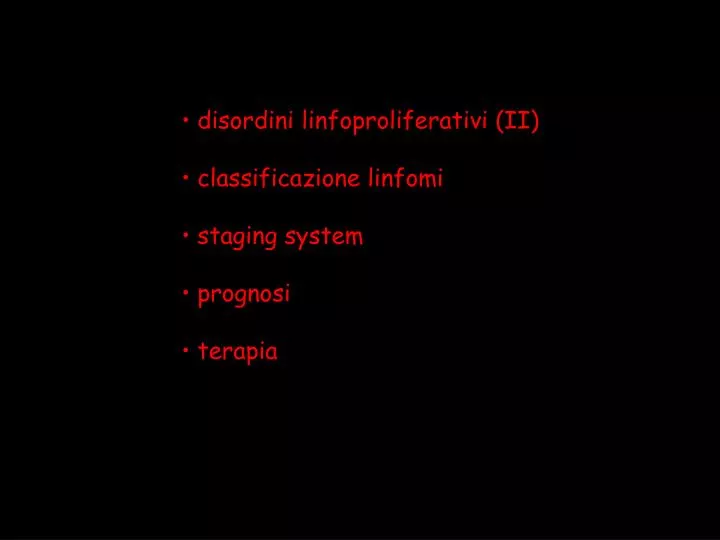
disordini linfoproliferativi (II) • classificazione linfomi • staging system • prognosi • terapia
B-Cell Neoplasms I. Precursor B-cell neoplasm: a. Precursor B-lymphoblastic leukemia/lymphoma II. Mature (peripheral) B-cell neoplasms a. B-cell chronic lymphocytic leukemia / small lymphocytic lymphoma b. B-cell prolymphocytic leukemia c. Lymphoplasmacytic lymphoma d. Splenic marginal zone B-cell lymphoma (+/- villous lymphocytes) e. Hairy cell leuekmia f. Plasma cell myeloma/plasmacytoma g. Extranodal marginal zone B-cell lymphoma of mucosa-associated lymphoid tissue type h. Nodal marginal zone lymphoma (+/- monocytoid B-cells) i. Follicle center lymphoma, follicular, j. Mantle cell lymphoma k. Diffuse large cell B-cell lymphoma • Mediastinal large B-cell lymphoma • Primary effusion lymphoma l. Burkitt's lymphoma/Burkitt's cell leukemia
T-Cell and Natural Killer Cell Neoplasms I. Precursor T cell neoplasm: a. Precursor T-lymphoblastic lymphoma/leukemia II. Mature (peripheral) T cell and NK-cell neoplasms a. T cell prolymphocytic leukemia b. T-cell granular lymphocytic leukemia c. Aggressive NK-Cell leukemia d. Adult T cell lymphoma/leukemia (HTLV1+) e. Extranodal NK/T-cell lymphoma, nasal type f. Enteropathy-type T-cell lymphoma g. Hepatosplenic gamma-delta T-cell lymphoma h. Subcutaneous panniculitis-like T-cell lymphoma i. Mycosis fungoides/Sézary's syndrome j. Anaplastic large cell lymphoma, T/null cell, primary cutaneous type k. Peripheral T cell lymphoma, not otherwise characterized l. Angioimmunoblastic T cell lymphoma m. Anaplastic large cell lymphoma, T/null cell, primary systemic type
NON-HODGKIN LYMPHOMAS: • 1) non-Hodkin lymphomas are a diverse collection of approximately • 40 entities, with different immunopathologic and cytogenetic characteristics • 2) the most frequent entities are the: • - Follicle Centre lymphoma (FCL) • - Diffuse Large Cell lymphoma (DLCL) • 3) B-cell derived are by far more frequent compared to T-cell derived • (90% vs. 10%)
INCIDENZA DEI VARI TIPI DI LINFOMA NON-HODGKIN 28% 16% 6% 30% 20% B-cell DLCL FCL T-cell NHL non follicular low grade NHL Other lymphomas
Hodgkin's lymphoma (Hodgkin's Disease) a.Nodular lymphocyte predominance Hodgkin's lymphoma b.Classical Hodgkin's lymphoma • Nodular sclerosis Hodgkin's lymphoma • Lymphocyte-rich classical Hodgkin's lymphoma • Mixed cellularity Hodgkin's lymphoma • Lymphocyte depletion Hodgkin's lymphoma
Hodgkin's Disease - Classification Type Histologic Features Frequency Prognosis Nodular sclerosis Bands of fibrosis, Most frequent type, Good Lacunar cells more common in women most are stage I-II Mixed cellular Composed of many Most frequent Fair different cells in older persons, most are stage III second most frequent overall Lymphocyte predominance Mostly B-cells and few Uncommon Good Reed-Sternberg variant cells most are stage I or II Lymphocyte depletion Many Reed-Sternberg Uncommon Poor cells and variants most are stage III or IV
CARATTERIZZAZIONE RISCHIO PROGNOSTICO: biopsia linfonodale biopsia osteo-midollare tipizzazione immunofenotipica tipizzazione molecolare stadiazione della malattia
DEFINIZIONE RISCHIO PROGNOSTICO: biopsia linfonodale biopsia osteo-midollare tipizzazione immunofenotipica tipizzazione molecolare stadiazione della malattia fattori di rischio
Fattori con valore prognostico sfavorevole indipendente: • performance status > 2 • LDH > normale • siti extranodali 2 • stadio III o IV • età > 60 anni • No. di fattori presenti • Tipo di rischio prognostico • 0-1 basso (L) • 2 intermedio-basso (LI) • 3 intermedio-alto (HI) • 4-5 alto (H)
Overall Survival in DLCL according to risk group defined by Age-Adjusted IPI (PS, stage, LDH) C R 5-yr Risk group Score Rate survival (%) (%) Low 0 92 83 Low-intermediate 1 78 69 High-intermediate 2 57 46 High 3 46 32
IIL prognostic system • Age (< vs. > 60 vs) • Sex (F vs M) • Extranodal sites (0-1 vs 2) • Serum LDH (normal vs elevated) • B symptoms (absent vs present) • ESR (less than 30 vs at least 30)
Federico M et al., Blood 2000, 95: 783-789 Prognosis of follicular lymphoma: a predictive model based on a retrospective analysis of 987 cases
Hodgkin's lymphoma (Hodgkin's Disease) a.Nodular lymphocyte predominance Hodgkin's lymphoma b.Classical Hodgkin's lymphoma • Nodular sclerosis Hodgkin's lymphoma • Lymphocyte-rich classical Hodgkin's lymphoma • Mixed cellularity Hodgkin's lymphoma • Lymphocyte depletion Hodgkin's lymphoma
Hodgkin's Disease - Classification Type Histologic Features Frequency Prognosis Nodular sclerosis Bands of fibrosis, Most frequent type, Good Lacunar cells more common in women most are stage I-II Mixed cellular Composed of many Most frequent Fair different cells in older persons, most are stage III second most frequent overall Lymphocyte predominance Mostly B-cells and few Uncommon Good Reed-Sternberg variant cells most are stage I or II Lymphocyte depletion Many Reed-Sternberg Uncommon Poor cells and variants most are stage III or IV
Factors with independent prognostic value for survival in lymphomas of both high and low grade histology Prognostic classification Factor age >60 performance status I P I serum LDH level (New Engl J Med 1993) Ann Arbor stage extranodal involvement Performance status aa I P I serum LDH level Ann Arbor stage (New Engl J Med 1993) Age (>60) Sex (male) I I L ESR () (Blood 2000) Serum LDH level () Systemic symptoms extranodal involvement
13-gene predictor: cured gene-espression signature fatal/refractory gene-espression signature
13-gene outcome predictor: IPI-outcome predictor:
13-gene predictor: cured gene-espression signature fatal/refractory gene-espression signature
Overall Survival of advanced-stage DLCLwith 3rd generation chemotherapy regimens
Corradini P et al, Blood 1997 Jan 15;89:724-31 Tarella C et al, Leukemia 2000 Apr 14:740-7 Turin-group experience with the i-HDS scheme a “high-dose” approach aimed to obtain maximal tumor cytoreduction and to exploit the in vivo-purging effect operated by chemotherapy
DHAP x 2 G - C S F G - C S F I-HDS SCHEME FOR HIGH-RISKFCL PATIENTS MTX8 g/sqm VP-162 g/sqm CTX7 g/sqm APO x 2 PBPC/BMharvest MITOX + L-PAM+ PBPC autograft DEX 40
100 90 80 70 60 % surviving 50 40 30 20 10 1 2 3 4 5 6 7 8 9 years I-HDS REGIMEN IN FCL: results of the Torino group experience Leukemia 2000, 14: 740-747 • CR RATE OF 79% • ACCEPTABLE RATE OF EARLY AND LATE TOXICITIES • A PROJECTED EFS AT 9 YEARS OF 62% AND A PROJECTED OS OF 78% 100 90 80 70 60 % surviving 50 40 30 Overall survival 20 Event-free survival 10 0 0 0 1 2 3 4 5 6 7 8 9 0 years
Gianni AM et al; NEJM 1997; 336: 1290-97“HDS vs MACOP-B in aggressive B-cell NHL“
DEVELOPMENT OF MONOCLONAL ANTIBODIES HUMAN MOUSE HUMANIZED CHIMERICAL
UNLABELED CHIMERIC ANTIBODY IMMUNOTOXIN RADIOCONJUGATE
Meccanismo d’azione mAbs • effetto diretto • signaling apoptosi • citossico (tossine o radiomarcati) • effetto indiretto • complemento • ADCC (NK, GN) • immunosensibilizzazione
Principali anticorpi monoclonali “unlabelled” STRUTTURA INDICAZIONI ANTIGEN NAME FCL,MCL,HCL, DLCL Trapianto LLC, Trapianto FCL Chimerico Chimerico umanizzato umanizzato umanizzato Rituximab Basiliximab Daclizumab Campath 1H Epratuzumab CD20 CD25 CD52 CD22
RADIOIMMUNOCONJUGATE Effector mechanisms Radiation-induced cytolysis • TARGET ANTIGENS: • NOT SHED • NOT INTERNALIZED ? NB. Properties of each immunoconiugate depend on which isotope is chosen
RITUXIMAB C-HDS + Rituximab schedule hd-Ara-C VP16 + CDDP hd-CY PBSC autografting A P O PBSC harvest PBSC harvest G-CSF G-CSF G-CSF
C-HDS + Rituximab in high-risk DLCL patients: a multicenter italian study CR OS 71 % (3yr.) R- HDS 82% historical 46-57% 32-46% (5yr.)
identification of residual disease The role of 67Ga scanning or FDG-PETin discriminating between active or fibrotic residual masses is well established
identification of residual disease The value of molecular biology techniques (PCR) in evaluating the minimal residual disease in patients with Bone Marrow involvement at presentation
100 PCR negative 80 60 % surviving 40 PCR positive P<0.005 20 0 2 4 6 8 0 10 12 years DFS comparison between PCR-positive and PCR-negative patients
IMMUNOTERAPIA NEI DISORDINI LINFOPROLIFERATIVI • percentuale di guarigione ancora insufficiente • crescita abbastanza lenta • markers tumore-specifici o lineage-specifici • chemioterapia efficace ma non eradicante • monitoraggio della malattia minima residua (MMR) • MMR spesso MDR+ • immunosensibilita’ della MMR • modelli animali disponibili
VACCINATION SCHEDULE week 0 2 6 10 14 24 28 Id/KLH (0.5 mg + 0.5 mg) GM-CSF (150 µg/sqm) Protein-based vaccine • 15 MM in first remission after HDS and PBPC infusion;