Download
1 / 38
380 likes | 668 Views
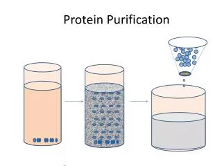
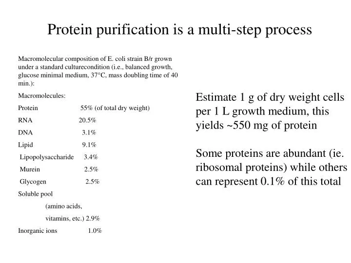
Protein purification is a multi-step process. Macromolecular composition of E. coli strain B/r grown under a standard culturecondition (i.e., balanced growth, glucose minimal medium, 37°C, mass doubling time of 40 min.): Macromolecules:

E N D
Protein purification is a multi-step process Macromolecular composition of E. coli strain B/r grown under a standard culturecondition (i.e., balanced growth, glucose minimal medium, 37°C, mass doubling time of 40 min.): Macromolecules: Protein 55% (of total dry weight) RNA 20.5% DNA 3.1% Lipid 9.1% Lipopolysaccharide 3.4% Murein 2.5% Glycogen 2.5% Soluble pool (amino acids, vitamins, etc.) 2.9% Inorganic ions 1.0% Estimate 1 g of dry weight cells per 1 L growth medium, this yields ~550 mg of protein Some proteins are abundant (ie. ribosomal proteins) while others can represent 0.1% of this total
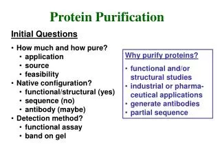
Proteins have unique properties resulting from their amino acid composition • Localization • Charge • Hydrophobicity • Size • Affinity for ligands Arbitrary protein
The charge on a protein is dependent upon pH • The content of amino acids with ionizable side chains determines the overall charge of a protein • Thus, a protein containing a majority of basic • residues (ie. R and K) will be positively charged • and will bind to a cation-exchange support Ion exchange column Supports (examples)
Cation exchange chromatography • Protein samples are applied to this column at low ionic strength, and positively charged proteins bind to the column support • Proteins are eluted using a gradient of increasing ionic strength, where counterions displace bound protein, changing pH will also elute protein • Choice of functional groups on distinct column supports allow a range of affinities Na+Cl- - - - Protein - - - - - - - - - - - - - - - - -
Conversely, at a pH two orders of magnitude above their pKa, acidic amino acids will be negatively charged, thus proteins with a majority of acidic amino acids (D and E) will be negatively charged at physiological pH • Negatively charged proteins can be separated using • anion exchange chromatography
Anion exchange chromatography • Protein samples are applied to this column at low ionic strength, and negatively charged proteins bind to the column support • Proteins are eluted using a gradient of increasing ionic strength, where counterions displace bound protein, changing pH will also elute protein • Choice of functional groups on distinct column supports allow a range of affinities • Bead size affects resolution in both anion and cation exchange Na+Cl- Protein + + + + + + + + + + + + + + + + + + + +
At a specific pH, the isoelectric point, a protein will not exhibit a charge
Hydrophobic Interaction Chromatography • Although most hydrophobic amino acids are buried in • the interior of proteins, many proteins have hydrophobic • surfaces or patches which can be used for separation • A protein’s hydrophobic character is typically enhanced • by addition of high salt concentrations • Proteins are eluted from HIC columns via a gradient of • high salt to low salt concentrations
Salt effects on protein solubility At low ionic strengths, the charges on the surface of a protein attract counter ions, decreasing electrostatic free energy and increasing solubility. Addition of low concentrations of salt, then, increase solubility of proteins ("salting in"). At high salt concentrations, however, protein solubility decreases ("salting out"). This is due to electrostatic repulsion between the surface ions and the hydrophobic interior of the protein and to the avid interaction of salts with water. This disrupts the ordered water in the hydration layer. Salts vary in their ability to salt out proteins and generally follow the Hofmeister series: Cations: NH4+ > K+ > Na+ > Mg++ > Ca++ > guanidium Anions: SO4-- > HPO4-- > acetate > citrate > tartrate > Cl- > NO3-
Proteins can be separated on the basis of size • Gradient centrifugation • Gel filtration
10% Glycerol 40% Glycerol Proteins can be separated by their sedimentation properties Function of both size and shape
Gel Filtration provides a molecular sieve Figures from Scopes, Protein Purification on Reserve
A protein’s substrate preference can be used in a very specific purification step Intrinsic If a protein binds ATP, put over a column support that has ATP crosslinked on it, thus selecting for ATP-binding proteins (can be done or a wide range of substrates such as sugars, Proteins, etc.) Added Specific protein domains can be fused to proteins of interest at the gene level to facilitate purification (ie. Fuse a maltose binding protein domain to any random protein, then it will bind specifically to a maltose containing column)
Metal chelation is a popular affinity purification method Various “expression vectors” create fusions to poly-Histidine tags, which allow the protein to bind to columns containing chelated metal supports (ie. Ni+2) Figures from Qiagen Product literature
Carbonic Anhydrase is a non-abundant protein in M. thermophila Taken from paper on reserve Increasing purity
Purification Analysis • Spectroscopy • Gel Electrophoresis • Enzymatic Assay
Spectroscopy is a study of the interaction of electromagnetic radiation with matter A = ecl Absorbance = extinction coefficient x concentration x path length Units: None = M-1 cm-1 M cm Beer-Lambert Law The amount of light absorbed is proportional to the number of molecules of the chromophore, through which the light passes
How do molecules absorb light? Photon energy causes electrons to jump to higher energy levels in molecular orbitals
Increased delocalization shifts the absorption band closer to the visible range
Tetrapyrroles (heme, chlorophyll) make proteins visible
c-type cytochromes have a characteristic absorbance spectrum Isobestic point
About one-third of all enzymes require one or more metal ions for catalytic activity
Some transition metals absorb in the visible range Several hyperthermophilic archaeal species have also been shown to be dependent on tungsten (W), also Cd important in diatoms
Proteins bind metals based on size, charge, and chemical nature Each metal has unique properties regarding ionic charge ionic radii, and ionization potential Typically, metals are classified as “hard” or “soft” in correlation with their ionic radii, electrostatics, and polarization Hard metals prefer hard ligands, soft prefer soft, Borderline metals can go either way.
Biological roles of transition metals (not just limited to proteins*) • Coordination • Structure (protein and protein-substrate) • Electrophilic catalysis • Positive charge attracts electrons, polarize • potential reactant, increase reactivity • General Acid – Base catalysis • Redox reactions • Metalloorganic chemistry • Free radicals *why did we add EDTA to lyse the outer membrane
Chelators bind metals tightly via multiple interactions These may be difficult to make out, to get the idea simply look at the heme figure
L L L L L M M L L L L L L L M M L L L L L L L Metals favor distinct coordination in proteins Tetrahedral Trigonal bipyramidal M = Metal L = Ligand Octahedral Square Planar
Transition metals are Lewis acids Acid-Base definitions Lowry-Bronsted – an acid is a substance that gives up a proton, and a base accepts a proton Lewis – an acid can take up an electron pair to form a covalent bond, while a base can furnish an electron pair Zinc acts as a Lewis acid to generate nucleophiles Nucleophiles are Lewis bases
Molecular Weight Markers Purified 20S Proteasome Protein’s migrate as a function of size using SDS-PAGE SDS is a strongly anionic detergent that associates with denatured polypeptides proportionally to their size, thus the charge on a protein becomes independent of it’s sequence but a function of it’s size, however, the sample must be completely denatured by heat and reductant prior to loading a subunit b subunit Good analysis for an “apparent” molecular weight, and subunit composition
Protein’s migrate as a function of charge and size under non-denaturing conditions Non-denaturing conditions allow you to examine the native active state of an enzyme (can stain proteins based on their activity) How many bands would you observe on a non-denaturing gel using purified proteasome? How would you determine the stoichiometry of oligomeric enzymes using this technique?
Gel electrophoresis can be used in preparative as well as analytical ways
Gel electrophoresis can also separate proteins by pH (Isoelectric focusing) Low pH High pH pI =7.5 pI=5.4 pI=6.8 - +
Anti-sera can be used to detect specific proteins Sufficiently separated proteins in an SDS-PAGE can be transferred to a solid membrane for Western Blot analysis. For this procedure, an electric current is applied to the gel so that the separated proteins transfer through the gel and onto the membrane in the same pattern as they separate on the SDS-PAGE. All sites on the membrane which do not contain blotted protein from the gel can then be non-specifically "blocked" so that antibody (serum) will not non-specifically bind to them, causing a false positive result.
Developing a Western Blot To detect antigen blotted on the membrane, a primary antibody (serum) is added at an appropriate dilution and incubated with the membrane. If there are any antibodies present which are directed against one or more of the blotted antigens, those antibodies will bind to the protein(s) while other antibodies will be washed away at the end of the incubation. In order to detect the antibodies which have bound, anti-immunoglobulin antibodies coupled to a reporter group such as the enzyme alkaline phosphatase are added (e.g. Goat anti-human IgG- alkaline phosphatase). This anti-Ig-enzyme is commonly called a "second antibody" or "conjugate". Finally after excess second antibody is washed free of the blot, substrate is added which will precipitate upon reaction with the conjugate resulting in a visible band where the primary antibody bound to the protein.
Enzymatic assays measure enzyme activity Change in something (Usually O. D.) Time Specific Activity vs. Total Activity