Download
1 / 29
300 likes | 541 Views
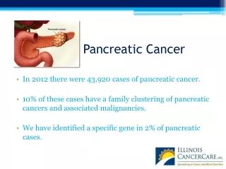
presenters: Grant Stearns, Robert Joung. Future Strategy for Pancreatic Cancer Screening. facilitator: Dr. Steve Potter. The Problem. A current screening strategy that diagnoses pancreatic cancer at a stage that offers resectability and treatment (pre-stage 2) is needed.

E N D
presenters: Grant Stearns, Robert Joung Future Strategy for Pancreatic Cancer Screening facilitator: Dr. Steve Potter
The Problem • A current screening strategy that diagnoses pancreatic cancer at a stage that offers resectability and treatment (pre-stage 2) is needed. • An accurate non-invasive technique is needed to justify more invasive techniques. • Maintain specificity while increasing sensitivity. AUC above 0.9 is considered highly accurate.
Analytic Methods • Researched Current Screening Methods. • Analyzed Current Screening Methods. (ROC Curves) • Researched Future Strategies. • Chose Future Strategy and Created Layout for Future Strategy. • Created Additive Value Model Risk Assessment Worksheet. • Refined Future Strategy.
Future Strategy Template [Iacobuzio-Donahue, C. A., S. Yachida, et al. 2010.]
Bob is an obese 52 year-old black male who has had chronic pancreatitis for less than two years and who also has one immediate family member that developed pancreatic cancer. Bob has 45 points on the Risk Assessment. This makes Bob a High Risk patient.
Many methods of testing Reverse Phase Protein Array (RPPA) Luminex Assays Forward phase or antibody array Multiplexed Bead Assays Decreasing cost Mass diagnosis Good ROC values Why biomarkers? [Li, C. et al. 2011] [Grote, T. et al. 2008] [http://www.oasiswebdevelopment.com/newrpci/services/multiplex.html][Ryozawa, S., H. Iwano, et al. 2011] [http://www.mcponline.org/content/4/4/346.full]
Biomarker Panels Comparison Biomarker Panel 1 Biomarker Panel 2 [Hoimes, C. et al. 2009] [Farrell, J. et al. 2009]
Combination of 3 mRNA biomarkers (ACVR1, DMXL2, DPM1) Differentiate between pancreatic cancer patients and all others Sensitivity of 93% and Specificity of 90% Addition of a bacterial biomarker (S. Mitis) to the panel Differentiates between subjects with pancreatic cancer, chronic pancreatitis, and healthy controls Sensitivity of 93% and a Specificity of 85% Step 2: Selected Biomarker Panel [Hoimes, C. et al. 2009]
Invasive Techniques Comparison [Glasbrenner, B. 2000.] [Kitano, M. 2011.] [Grenacher, L. 2004.] [Li, K. C. 1988] [Pennant, M. 2010.]
Invasive Techniques Comparison CT EUS - ERCP EUS - FNA Sensitivity: 94.76% Specificity: 89.165% AUC: 0.9196 Sensitivity: 92% Specificity: 85% AUC: 0.885 Sensitivity: 86.5% Specificity: 87.5% AUC: 0.87 EUS MRI PET Sensitivity: 80.738% Specificity: 89.119% AUC: 0.85 Sensitivity: 89.375% Specificity: 86.725% AUC: 0.8805 Sensitivity: 89.5% Specificity: 91.5% AUC: 0.905 [Glasbrenner, B. 2000.] [Kitano, M. 2011.] [Grenacher, L. 2004.] [Li, K. C. 1988] [Pennant, M. 2010.]
Advantages SN of 86.5% SE of 87.5% AUC of 0.87 Cost $2,440-$3,300 Access Risk Selected Invasive Technique EUS guided FNA [Glasbrenner,B. et al ; Kitano, M. et al.] [Ashida, R., Y. Arisaka, et al. 2011] [Ryozawa, S., H. Iwano, et al. 2011] [ Powis,M. et al. 2000.]
Incorporate the use of the AVM Risk Assessment Sheet to determine the risk level of a patient. Incorporate the use of the salivary biomarker panel to further assess the overall risk of a patient post-risk assessment. Incorporate the use of EUS-FNA, after a positive biomarker test, specifically to stage and identify the location of pre-stage 2 pancreatic cancer. Recommendations
Hassan, M. M., Bondy, M. L., Wolff, R. A., Abbruzzese, J. L., Vauthey, J.-N., Pisters, P. W., . . . Li, D. (2007). Risk factors for pancreatic cancer: case-control study. The American Journal Of Gastroenterology, 102(12), 2696-2707. Zavoral, M., Minarikova, P., Zavada, F., Salek, C., & Minarik, M. (2011). Molecular biology of pancreatic cancer. World Journal Of Gastroenterology: WJG, 17(24), 2897-2908. Institute, N. C. (2011). SEER Stat Fact Sheets: Pancreas Retrieved September 11, 2011, from http://seer.cancer.gov/statfacts/html/pancreas.html B. Glasbrenner, M. Schwarz, S. Pauls, G. Preclik, H. G. Beger, and G. Adler, "Prospective comparison of endoscopic ultrasound and endoscopic retrograde cholangiopancreatography in the preoperative assessment of masses in the pancreatic head," Digestive Surgery, vol. 17, pp. 468-474, 2000. Kitano, M., Sakamoto, H., Komaki, T., & Kudo, M. (2011). New techniques and future perspective of EUS for the differential diagnosis of pancreatic malignancies: contrast harmonic imaging. Digestive Endoscopy: Official Journal Of The Japan Gastroenterological Endoscopy Society, 23 Suppl 1, 46-50. doi: 10.1111/j.1443-1661.2011.01146.x Grenacher, L., Klauss, M., Dukic, L., Delorme, S., Knaebel, H. P., Düx, M., . . . Richter, G. M. (2004). [Diagnosis and staging of pancreatic carcinoma: MRI versus multislice-CT -- a prospective study]. Röfo: Fortschritte Auf Dem Gebiete Der Röntgenstrahlen Und Der Nuklearmedizin, 176(11), 1624-1633. Li, K. C., & Poon, P. Y. (1988). Sensitivity and specificity of MRI in detecting malignant spinal cord compression and in distinguishing malignant from benign compression fractures of vertebrae. Magnetic Resonance Imaging, 6(5), 547-556. Pennant, M., Takwoingi, Y., Pennant, L., Davenport, C., Fry-Smith, A., Eisinga, A., . . . Hyde, C. (2010). A systematic review of positron emission tomography (PET) and positron emission tomography/computed tomography (PET/CT) for the diagnosis of breast cancer recurrence. Health Technology Assessment (Winchester, England), 14(50), 1-103. Ashida, R., Y. Arisaka, et al. (2011). "The role of linear array EUS for diagnosis of pancreatic malignancies in the current situation." Digestive Endoscopy: Official Journal Of The Japan Gastroenterological Endoscopy Society 23 Suppl 1: 12-16. Ryozawa, S., H. Iwano, et al. (2011). "Genetic diagnosis of pancreatic cancer using specimens obtained by EUS-FNA." Digestive Endoscopy: Official Journal Of The Japan Gastroenterological Endoscopy Society 23 Suppl 1: 43-45. Kapke, G., & Stoddard, J. J. (2008). Biomarkers – boon or bane for researchers? Retrieved September 14, 2011, from http://www.covancecareers.com/biomarkers/pdf/boonorbane.pdf Citations
Chang, M. E. P. a. K. J. (2000). Endoscopic Ultrasound in the Clinical Staging and Management of Pancreatic Cancer: Its Impact on Cost of Treatment. CANCER CONTROL: JOURNAL OF THE MOFFITT CANCER CENTER, 7(5). • Li, C., Zolotarevsky, E., Thompson, I., Anderson, M. A., Simeone, D. M., Casper, J. M., . . . Lubman, D. M. (2011). A multiplexed bead assay for profiling glycosylation patterns on serum protein biomarkers of pancreatic cancer. Electrophoresis, 32(15), 2028-2035. doi: 10.1002/elps.201000693 • Grote, T., Siwak, D. R., Fritsche, H. A., Joy, C., Mills, G. B., Simeone, D., . . . Logsdon, C. D. (2008). Validation of reverse phase protein array for practical screening of potential biomarkers in serum and plasma: accurate detection of CA19-9 levels in pancreatic cancer. Proteomics, 8(15), 3051-3060. • Farrell, J. J., Zhang, L., Sugimoto, M., Hirayama, A., Soga, T., Zhou, H., . . . Wong, D. T. (2009). Multiple Salivary Biomarkers for Pancreatic Cancer Detection. [Meeting Abstract]. Gastroenterology, 136(5), A147-A147. • Iacobuzio-Donahue, C. A., S. Yachida, et al. (2010). "Distant metastasis occurs late during the genetic evolution of pancreatic cancer." Nature 467(7319): 1114-U1126. • Hoimes, C. J., Moyer, M. T., & Saif, M. W. (2009). Biomarkers for early detection and screening in pancreatic cancer. Highlights from the 45th ASCO annual meeting. Orlando, FL, USA. May 29-June 2, 2009. JOP: Journal Of The Pancreas, 10(4), 352-356. • Fishman, K. S. V. C. E. K. Y. L. D. (2005). Use of Reverse Phase Protein Microarrays and Reference Standard Development for Molecular Network Analysis of Metastatic Ovarian Carcinoma. Molecular and Cellular Proteomics, 346-355. • *Title slide photo credit: Adapted from Pancreatic Cancer Cell, SEM , Steve Gschmeissner /Science Photo Library
(Question) Specifics: Risk Assessment * Family History should include pancreatic cancer as well as breast cancer and colon cancer. ** A secondary relative is defined as a patient's aunts, uncles, and first cousins. *** A primary relative is defined as a patient's parents, grandparents, and siblings. **** Having an age less than 20 years puts the patient into the No Risk category, and all other points are void. ***** A light smoker is defined as a patient who has smoked for less than 20 years. ****** A heavy smoker is defined as a patient who has smoked for more than 20 years. [Hassan, M. M. et al. 2007] [Zavoral, M. et al. 2011],[Institute, N. C. 2011]
(Question) Risk Assessment Creation 1. Determined weighting of points with Additive Value Model. • Rank Risk Factors1,3 • Assign point values to risk factors. Give 100 pts to Rank 1 and Rank 2 onward will decend by a value equal to 100/ # of ranks. • Weighting will be: ( Rank point value ) / (Sum of all points ) • Adding the weighting values should give you a value of 1.0 if done correctly. 2. The highest risk characteristic from each risk factor was assigned that risk factor's weighting multiplied by 100. 3. The lowest risk characteristic from each risk factor was assigned the value of zero. Assigning a point value for all risk factors would make the point system less accurate and meaningful. 4. The intermediate characteristics within each risk value was assigned a point value based on NCI and research statistics relative to the highest risk characteristic. Hypothetically, if the highest risk characteristic's value were to be 100 points, and the statistic for that risk characteristic was 50%, then the second characteristic whose statistical value is 25% would be assigned a value of 50 points.2 5. The point scale was designed to set the highest possible risk at 100 points and the lowest possible risk at 0 points. 6. The Low Risk cut-off point for testing was determined to be 15 points based on the highest lowest value that one could have with only the characteristics of gender, race, the lowest age, and one secondary relative with a history of the relevant cancers. 7. The Low Risk/ High Risk transition point of 40 points was determined using a peer-reviewed research article's assessments of risk levels for patients as well as an adjustment for potential under-estimation of certain risks. [Hassan, M. M. et al. 2007], [Zavoral, M. et al. 2011],[Institute, N. C. 2011].
(Question) How often should a patient have a risk assessment? First Assessment: Age 20 Future assessments should be determined by the doctor. Guidelines: - Re-assessment every 2 years at the least. - If the patient has more than 10 points, there should be a re-assessment at every annual checkup. - If the patient is categorized as Low Risk and has Type-II Diabetes or Pancreatitis, the decision of future testing should be left to the patient's physician. *All values based off of Risk Assessment Chart.
(Question) Stages and rates • Less than 5% of patients survive 5 years • In stage I, 21.5% of patients survive 5 years • In stage II, 8.6% of patients survive 5 years • In stages III and IV combined, 1.8% of patients survive 5 years • In unknown stages, 4.2% of patients survive¹ • The median survival rate is between 8 and 14 months² 1National Institute of Cancer, http://seer.cancer.gov/statfacts/html/pancreas.html 2Mahadevan A. et Al., Induction Gemcitabine and Stereotactic Body Radiotherapy for Locally Advanced Nonmetastatic Pancreas Cancer
(Question) Stages of Pancreatic Cancer • Stage 0: abnormal cells are found in the lining of the pancreas. • Stage I: cancer has formed in pancreas. -Stage Ia: The tumor is 2 cm or smaller -Stage Ib: The tumor is larger than 2 cm • Stage II: The cancer has spread. -Stage IIa: The cancer had spread to nearby tissues and organs -Stage IIb: The cancer has spread to lymph nodes • Stage III: The cancer has spread to the major blood vessels near the pancreas. • Stage IV: The cancer may be of any size and may have spread to distant organs (liver, lung and, peritoneal cavity). The cancer has metastasized. National Institute of Cancer, http://seer.cancer.gov/statfacts/html/pancreas.html
(Question) Fine Needle Aspiration • Fine Needle Aspiration Biopsy, Fine needle Aspiration Cytology • Cost: about $2,000 (With EUS/ Lung lesion) • Interpretation: Dyed sample tissues or cells. • Limit: Anesthesia might be needed. Most patients feel pain during the procedure. • Risk: The disease can be spread out through the needle tip. 1Everyday Health (http://www.everydayhealth.com/pancreatic-cancer/biopsy-and-staging.aspx)2AFH library. (http://www.alkalizeforhealth.net/Lneedlebiopsy.htm) 3W.E. Khalbuss,3rd KAMC CYTOPATHOLOGY SYMPOSIUM (http://www.ngha.med.sa/English/MedicalCities/AlRiyadh/Centers/Lab/Symposiums/Documents/Day2/FNA7.EBUS.EUS.pdf )
Cost: $900-$1800 Invasiveness: Use of an IV for contrast agent. Limitation: Cannot be used on obese patients and not recommended for pregnant women. (Question) Current Technique - CT Sensitivity: 94.76% Specificity: 89.165% AUC: 0.9196 Grenacher, L. et al. "[Diagnosis and staging of pancreatic carcinoma: MRI versus multislice-CT -- a prospective study]"
cost: [EUS] $500 [ERCP] $740 Invasiveness: Very Invasive Limitation:[EUS] limited diagnostic [ERCP] Iodine allergy. Empty stomach Risk: [ERCP] Complications. (Question) Current Technique - EUS and ERCP Sensitivity: 92% Specificity: 85% AUC: 0.885 Glasbrenner,B. et al. "Prospective comparison of endoscopic ultrasound and endoscopic retrograde cholangiopancreatography in the preoperative assessment of masses in the pancreatic head"
cost: $2,440-$3,300 Invasiveness: Very Invasive Limitation:[EUS] limited diagnoses risk:[EUS] allergic reaction [FNA] bruising. (Question) Current Technique - EUS and FNA Sensitivity: 86.5% Specificity: 87.5% AUC: 0.87 [Glasbrenner,B. et al] ; [Kitano, M. et al.]; [J Gastroenterol 2004]; [Digestive Endoscopy 201]
Cost: around $500 Invasiveness: Very Invasive Limitation: Limited diagnostic Risk: allergic reaction (Question) Current Technique - EUS Glasbrenner,B. et al "Prospective comparison of endoscopic ultrasound and endoscopic retrograde cholangiopancreatography in the preoperative assessment of masses in the pancreatic head" Kitano, M. et al. "New techniques and future perspective of EUS for the differential diagnosis of pancreatic malignancies: contrast harmonic imaging," Sensitivity: 80.738% Specificity: 89.119% AUC: 0.85
Cost: $400 - $3000 Invasiveness: None Invasive Limitation:Sensitive to motion Risk: cannot be used if ferromagnetic materials are in the body. Can cause skin irritation. (Question) Current Technique - MRI Grenacher, L. et al. "[Diagnosis and staging of pancreatic carcinoma: MRI versus multislice-CT -- a prospective study]" Li, K. C. et al. "Sensitivity and specificity of MRI in detecting malignant spinal cord compression and in distinguishing malignant from benign compression fractures of vertebrae" Sensitivity: 89.375% Specificity: 86.725% AUC: 0.8805
Cost: [CT] $1800, [MRI] $400-$3000 Invasiveness: non invasive Limitation: The image might not be accurate if certain metal is involved in the body. Risk:[CT] [MRI] No metal, can cause skin irritation (Question) Current Technique - CT and MRI Sensitivity: 90% Specificity: 100% AUC: 0.95 Fusari, M. et al. "Comparison between multislice CT and MR imaging in the diagnostic evaluation of patients with pancreatic masses"
Cost: $3,000-6,000 Limitation: Is not as good as other methods at staging and testing for resectability. Risk: the exposure to radiation. (Question) Current Technique - PET Sensitivity: 89.5% Specificity: 91.5% AUC: 0.905 Pennant, M. et al. "A systematic review of positron emission tomography (PET) and positron emission tomography/computed tomography (PET/CT) for the diagnosis of breast cancer recurrence"