Download
1 / 73
870 likes | 1.35k Views
Imaging of cerebral ischemia. John-Henry Corbett Department of clinical imaging science University of the Free State 3/08/2012. Stroke is a clinical term applied to any abrupt non-traumatic brain insult Infarction 75% Haemorrhage 25%

E N D
Imaging of cerebral ischemia John-Henry Corbett Department of clinical imaging science University of the Free State 3/08/2012
Stroke is a clinical term applied to any abrupt non-traumatic brain insult • Infarction 75% • Haemorrhage 25% • Treatment of ischemic stroke in the past has been largely preventative and supportive • Approval of thrombolysisfor acute stroke and development of neuroprotective drugs • Led to importance of rapid imaging and intervention in stroke management
Pathophysiology of brain damage after infarction • 1. Sodium-Potassium pump • No blood supply -> depletion of oxygen & glucose -> decreased ATP at cellular level • Sodium-Potassium pump cannot function • Usually 3 Na+ ions out of cell and 2 K+ ions in • Causes passive diffusion of Na+ to inside of cells, along with water • Leads to cytotoxic oedema • Gray matter more metabolically active
Pathophysiology of brain damage after infarction • 2. Calcium pump • Depolirazation of cells leads to release of toxic amino acids, especially Glutamate • Directly neurotoxic • Also opens calcium channels • Large influx of Ca2+ into cells • Damages intracellular organelles, especially mitochondria • Inflammation, necrosis and apoptosis results
Pathophysiology of brain damage after infarction • 3. Blood-Brain barrier • Damaged by combination of hypoxic damage to vascular endothelium, toxic damage of inflammatory molecules and free radicals • Mainly seen after reperfusion • Leads to vasogenicedema, inflammation and haemorrhagic transformation
Stages of stroke : Pathology and Imaging Findings • Hyperacute stage : < 12 hours • Imaging findings due to • cytotoxicedema • Early CT signs – can take up to 8 hours • DWI & ADC (MRI) – within minutes of onset • { Hypodense areas on CT & DWI/ADC edema represents core infarct } • thrombus in vessels • Hyperdense MCA or MCA dot sign on noncontrast CT • MRI : Loss of arterial flow voids on T2, low signal on T2* or high signal on FLAIR • If < 6 hours since symptom onset, CT perfusion or MRI perfusion important to detect penumbra
Stages of stroke : Pathology and Imaging Findings • Acute stage: 12 - 24 hours • Increase in cytotoxicedema and intracellular Ca2+ • Free radicals and enzyme activation leads to cell membrane damage and cell death • Increased tissue water • Increase T2 signal (6-8h ; 90% by 24h) and decreased T1 • Effacement of convexity of sulci and mild swelling of gyri, without mass effect
Stages of stroke : Pathology and Imaging Findings • Subacute stage: 2 days – 2 weeks • Blood-brain barrier breakdown • Rupture of swollen cells • = increase in extracellular fluid • Vasogenicedema • Maximum by 48-72 hours • Imaging • Increased edema with mass effect / herniation • Gyral and parenchymal enhancement • Haemorrhagic transformation
Stages of stroke : Pathology and Imaging Findings • Chronic Stage : 2 weeks – 2 months • Restoration of BBB • Resolution of vasogenicedema • Cleaning up of necrotic tissue • Imaging • Brain atrophy, gliosis, cavity formation • Ex vacuo dilatation of adjacent ventricle • Calcification and deposition of blood products may remain ( T2, GRE) • Wallerian degeneration
Imaging approach to acute stroke • Four P’s • Parenchyma • Assess early signs of acute stroke • Rule out haemorrhage • Pipes • Assess extracranial circulation (carotid & vertebral) and intracranial circulation for evidence of intravascular thrombus • Perfusion • Assess Cerebral blood volume (CBV), Cerebral blood flow (CBF) and Mean transit time (MTT) • Penumbra • Assess tissue at risk of dying if ischemia continues without recanalization of intravascular thrombus
Penumbra • Brain tissue is very sensitive to ischemia • Unlike muscle, there are no neuronal energy stores • In complete absence of blood flow, neuronal viability can only be maintained for 2-3 minutes • In acute stroke, ischemia is usually incomplete • Injured area of brain receiving collateral blood supply from uninured arterial and leptomeningeal territories • Acute cerebral ischemia may result in a central irreversibly infarcted tissue core, surrounded by a peripheral region of stunned cells that is called a penumbra • This region is potentially salvageable with early recanalization
Imaging Modalities • CT • Unenhanced CT • CT angiography • CT perfusion imaging • MRI • Conventional MR imaging • MR angiography • Diffusion-weighted MR imaging • Perfusion-weighted MR imaging
Role of CT in Acute stroke –Unenhanced CT • Widely available, fast & no IV contrast • Can identify haemorrhage • Contraindication to thrombolytic therapy • Can identify early stage acute ischemia • Due to increased water content in infarcted area • Early signs of stroke on CT (may only be seen > 8 hours) • Loss of insular ribbon • Obscuration of lentiform nucleus • Loss of gray-white matter differentiation • Sulcal effacement • Indirect sign • Hyerpdense middle cerebral artery (MCA) sign • Thromboembolism in M1 segment • Associated poor clinical outcome • Large territorial infarction and increased bleeding risk • MCA dot sign • Hyperdensity in M2 / M3 segments in Sylvian fissure
Hyperdense MCA sign MCA dot sign Obscuration of lentiform nucleus Insular ribbon sign
Role of CT in Acute stroke –Unenhanced CT • Importance of window setting • Detection of acute ischemic stroke on unenhanced CT images may be improved by using variable window width and center level settings • Standard W 80 L 20 to W 8 L 32 • Accentuates contrast between normal and oedematous tissue
Role of CT in Acute stroke –Unenhanced CT • Quantification of ischemic involvement • Involvement of > 1/3rd of MCA territory depicted on unenhanced CT, criterion for exclusion of patients from thrombolytic therapy due to potential increase in haemorrhage risk • European Cooperative Acute Stroke Study trial • ASPECTS • Alberta Stroke Program Early CT Score (2001) • 10 point topographic scoring system of MCA territory • Normal MCA territory is assigned 10 points • One point deducted for each area involved on unenhanced CT
As ASPECTS decrease, the probability of dependence, death and symptomatic hemorrhage increase
Involvement of M1 region, insular cortex (I) and lentiform nucleus (L) ASPECTS score of 7
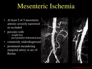
Role of CT in Acute stroke – CT angiography • Can assess intracranial and extracranial circulation • Coverage from aortic arch to the circle of Willis • Intra arterial thrombolysis may be more effective than IV in patients with acute stroke and significant thrombus burden • CT angiography can thus help guide appropriate treatment if it demostrates high thrombus burden • Can help identify cause of ischemic event (carotid & vertebral) • Visualization of arch and great vessels aids guidance for the interventional neuroradiologist
Hyperdense left MCA. Intravascular thrombus in this segment on CT angiography.
Role of CT in Acute stroke – CT perfusion imaging • Measure the following perfusion parameters • Cerebral Blood Volume (CBV) • Volume of blood per unit of brain tissue • Normal 4-5 ml / 100g • Cerebral Blood Flow (CBF) • Volume of blood flow per unit of brain tissue per minute • Normal for gray matter = 50-60 ml / 100g / min • Mean Transit Time (MTT) • Time difference between arterial inflow and venous outflow • Time to peak enhancement
Role of CT in Acute stroke – CT perfusion imaging • CBF = CBV / MTT • Perfusion features of penumbra • ↑ MTT , ↓ CBF, ↔ CBV (↑) • Perfusion features of infarcted tissue • ↑ MTT , ↓ CBF, ↓ CBV
Role of CT in Acute stroke – CT perfusion imaging • MTT perfusion map is the most sensitive indicator of stroke • CBF and CBV more spicific in distinguishing ischemia from infarction MTT CBF CBV
Role of CT in Acute stroke – CT perfusion imaging • Pitfalls • Slice selection • Large volume of brain tissue can be excluded eg posterior fossa & brainstem • Chronic white matter changes & old infarcts • especially if severe and asymmetrical (–> look at pre-contrast) • Arterial vascular stenosis • Extracranial carotid stenosis, intracranial carotid stenosis or proximal cerebral artery stenosis (-> importance of CT angio) • Vasospasm • Seizure mimicking stroke • Increased perfusion of ictal regions, suggesting ischemia in contralateral hemisphere
Role of MRI in Acute stroke – Conventional MR imaging • T1 & T2 spin echo, FLAIR, T2* weighted gradient echo, T1 post gadolinium • More sensitive and specific than CT in the first few hours after stroke • MRI findings in hyperacute cerebral ischemia • 1. High signal in white matter on T2 and FLAIR • 2. Loss of gray-white matter differentiation • 3. Sulcal effacement and mass effect
Role of MRI in Acute stroke – Conventional MR imaging • 4. Loss of arterial flow voids on T2, low signal on T2* or high signal on FLAIR – intravascular thrombus • 5. Acute intracranial haemorrhage seen as area of blooming on T2* T2 FLAIR Gradient echo
Role of MRI in Acute stroke – MR Angiography • Evaluation of intravascular thrombus in cerebral vessels • Evaluation of carotid bifurcations • Time of flight angiography or contrast enhanced Left proximal MCA thrombus Basilar artery throbus
Role of MRI in Acute stroke – Diffusion-weighted MR imaging • Areas of cytotoxicedema(restricted motion of water molecules) appear bright on diffusion weigted images • As early as 30 minutes after onset of ischemia • High signal up to 5 days • Mildly increased signal 1-4 wks • ADC Map • Decreased from 30 minutes after onset to 5 days • Then increases and reaches normal in 1-4 weeks • Likely due to development of vasogenicedema with cytoxicedema
Role of MRI in Acute stroke – Diffusion-weighted MR imaging • After weeks / months, gliosis develops • Increased extracellular water • DWI cannot be used alone to determine infarct age – compare DWI & ADC • Tissue with reduced ADC value almost always undergoe irreversible infarction • Ischemic regions that are still viable, may appear normal on initial DWI (↑ MTT , ↓ CBF, ↔ CBV) • Early findings of normal DWI and altered perfusion parameters may indicate tissue at risk - Penumbra
DWI ADC Posterior circulation acute ischemic stroke
ADC DWI Old infarcts right frontal and left parietal
Role of MRI in Acute stroke – Perfusion-weighted MR imaging • DWI – identifies areas of irreversibly infarcted tissue • PWI – identifies areas of reversible ischemia as well • Endogenous techniques – arterial spin labeling • Exogenous techniques – gadolinium • Similar perfusion maps to CT • MTT, CBV & CBF • In acute stroke, region showing diffusion and perfusion abnormalities = core infarct • Normal DWI and abnormal PWI = Penumbra
MRI -DWI MRI - PWI Precontrast CT Left MCA infarction. Match in DWI and PWI, so no penumbra. Therefore not candidate for thrombolytic therapy.
CT vs MRI in acute stroke • CT is widely available and fast • Noncontrast CT, CT angiography and CT perfusion can be performed in under 15 minutes • Superior to MRA in evaluating the vessels • Less artefacts and better quantification of lesions • MRI stroke protocol takes longer • Conventional MRI, DWI, MRA and PWI • No radiation • Can be performed without contrast • Arterial spin labelling, time-of-flight • The two modalities are equally useful for evaluating acute stroke • Equivalent depiction of the penumbra
References • Srinivasan A, Mayank G, Faisal A et al. State of the art imaging of acute stroke. Radiographics 2006 ; 26: S75-S95. • Allmendinger A, Tang E, Lui Y, et al. Imaging of Stroke: Part 1, Perfusion CT – Overview of technique, Interpretation Pearls and Common Pitfalls. American Journal of Roentgenology2012 ; 198: 52-62. • Kanekar S, Zacharia T, Roller R. Imaging of Stroke : Part 2, Pathophysiology at the Molecular and Cellular Levels and Corresponding Imaging Changes. American Journal of Roentgenology2012 ; 198: 63-74. • Best A, Acosta N, Fraser J, et al. Recognizing False Ischemic Penumbras in CT Brain Perfusion Studies. Radiographics 2012; 32: 1179-1196. • Leiva-Salinas C, Provenzale M, Wintermark M. Responses to the 10 Most Frequently Asked Questions About Perfusion CT. American Journal of Roentgenology2011 ; 196:53-60