Download
1 / 28
290 likes | 508 Views
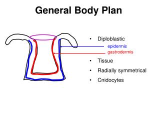
Basic embryonic body plan. I. Ectoderm . 60- hour chick embryo 15x. 60 X. Neural crest development. Neural crest again. Mesoderm . Neural tube 3. somite 5. yolk sac 7. aorta Amniotic cavity 4. nephrogenic cord 6. notochord 8. embryonic coelom.

E N D
Basic embryonic body plan I. Ectoderm
Neural tube 3. somite 5. yolk sac 7. aorta Amniotic cavity 4. nephrogenic cord 6. notochord 8. embryonic coelom
Development of the Heart • Splanchnic mesoderm : an area of the endoderm induces development of the cardiogenic mesoderm ; BMP, FGF • Heart and great vessels form from bilaterally paired tubes that fuse in the midline beneath the foregut to produce a single tube • See p. 118, Figure 6-15 • Layers of the single tube: • endocardial lining • “cardiac jelly” • Myocardium • Epicardium
The straight tubular heart • Endocardial tubes fuse one tube • Common cardinal veins flow into R and L sinus venosus • Sinus venosus leads to atrium • Atrium leads to ventricle • Outflow tract = truncusarteriosus
D-loop-- S-shape • See p. 117, Figure 6-14.
Blood and Blood Vessels • Blood islands form in mesodermal wall of yolk sac ( note: extraembryonic ) • Blood islands contain hemangioblasts • Inducer may be signaling molecule Indian hedgehog • Central part of blood islands hemocytoblasts • Peripheral cells endothelial cells lining the vessels.
Development of the Endoderm • See Figure 6-20, p. 122 • Three divisions: Foregut, Midgut, Hindgut • FGF-4 influences devel. of the hindgut • Hox genes + retinoic acid regulates regional development along the digestive tract • 1) Flat intraembryonic sheet of endoderm • 2) Lateral body folds form that sheet into a tubular structure • 3) Head and Tail folds form foregut, hindgut
Hox genes determine the form, number, and evolution of repeating parts, such as the number and type of vertebrae in animals with backbones. In the developing chick (left), the Hoxc-6 gene controls the pattern of the seven thoracic vertebrae (highlighted in purple), all of which develop ribs. In the garter snake (right), the region controlled by the Hoxc-6 gene (purple) is expanded dramatically forward to the head and rearward to the cloaca.