Download
1 / 1
10 likes | 187 Views
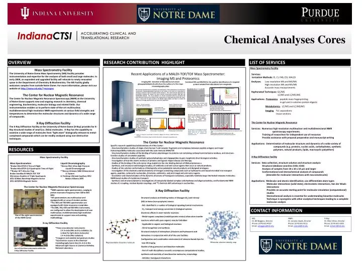
Representative bioactive molecule. One of the three instruments within the X-Ray Diffraction Facility. Molecular electron density map. Chemical Analyses Cores. OVERVIEW. RESEARCH CONTRIBUTION HIGHLIGHT. LIST OF SERVICES. Mass Spectrometry Facility Services:

E N D
Representative bioactive molecule One of the three instruments within the X-Ray Diffraction Facility Molecular electron density map Chemical Analyses Cores OVERVIEW RESEARCH CONTRIBUTION HIGHLIGHT LIST OF SERVICES Mass Spectrometry Facility Services: Ionization Methods: EI, CI, FAB, ESI, MALDI Analyses: Low resolution MS and MS/MS High resolution MS and MS/MS Accurate mass measurements Hyphenated Techniques: GC/MS LC/MS and LC/MS/MS Applications: Proteomicspeptide mass fingerprinting in-gel and in-solution protein digests MetabolomicsLC/MS and LC/MS/MS ImagingTLC separations tissue sections Mass Spectrometry Facility The University of Notre Dame Mass Spectrometry (MS) Facility provides instrumentation and expertise for the analyses of both small and large molecules. In early 2009, an expanded and upgraded facility will relocate to newly renovated space in the Department of Chemistry & Biochemistry. The MS Facility gladly welcomes samples from outside Notre Dame. For more information, please visit our website athttp://www.nd.edu/~massspec The Center for Nuclear Magnetic Resonance The Center for Nuclear Magnetic Resonance Spectroscopy (NMR) at the University of Notre Dame supports new and ongoing research in chemistry, chemical engineering, biochemistry, molecular biology and related fields. Our instrumentation enables us to perform state-of-the-art multinuclear, multidimensional high resolution NMR experiments at various field strengths and temperatures to determine the molecular structures and dynamics of a wide range of compounds. X-Ray Diffraction Facility The X-Ray Diffraction Facility at the University of Notre Dame (X-Ray) provides for X-Ray structural studies of small (ca. 2kDa) molecules. X-Ray has the capability to examine a wide range of molecules from “light atom” biologically relevant to metal-complexed compounds which can be readily analyzed using non-destructive techniques. • Structural analysis and binding modes of dioxygen (O2) and nitrosyl • (NO) in heme (iron-porphyrin) mimics • - NO: identified in a number of biological signaling/control mechanisms • - O2: transport and energy conversion in biological systems • Electronic effects in atom transfer reactions • - Metal-organic complexes (metalloprotein mimics) allow atom transfer • reactions which with pure organics may be forbidden • - Applicable to organic and biological reactions • - Chiral recognition and synthesis • Structural analysis of siderophore, β-lactam and hydroxamic acid • derivatives in conjunction with all of the core facilities • Configuration and coordination environment of rotaxane-based dyes for • near-IR imaging • Studies of drug precursor and bioactive molecules • - Part of multi-disciplinary research; encompasses computational studies, • synthesis and reactivity of new bioactive molecules, enzymology • - Inhibitor, host/guest interactions Imaging MS: detection of Mycobacterium avium glycopeptidolipids directly from the surface of a thin-layer chromatography plate We recently reported (1) that acetylation and methylation of the 6-deoxytalose and rhamnose constituents of glycopeptidolipids expressed on the surface of M. avium are responsible for eliciting an immune response through toll-like receptor 2. This study involved substantial TLC purification followed by analysis via direct infusion electrospray ionization (ESI) MS and ESI/MS/MS methods. The recent addition of a Bruker AutoflexIII (left) has permitted the direct detection from the TLC surface of the components of a total lipid extract from M. avium. Figure 1 shows the TLC plate loaded on the MALDI target adapter, and Figure 2 reveals the molecular ion image obtained from one lane of the separation. Recent Applications of a MALDI-TOF/TOF Mass Spectrometer: Imaging MS and Proteomics Combined MS and MS/MS for the positive identification of a targeted protein isolated from Mycobacterium smegmatis MASCOT Search Results Gel band “3” digest PROTEIN IDENTIFIED: gi|118467544 Mass: 29903 Score: 106 Expect: 5.9e-006 Queries matched: 7 tetrahydrofolate dehydrogenase/cyclohydrolase FolD [Mycobacterium smegmatis str. MC2 155] Figure 1. TLC plate mounted in MALDI-TOF target plate adapter Figure 2. Molecular ion image showing different m/z regions represented by various colors. acquire MALDI-TOF mass spectra at multiple points along the lane Figure 1. Barely visible Coomassie blue stained gel band which was excised, destained, and digested with trypsin to yield peptide fragments for MS analysis. Figure 2. Results of peptide mass fingerprint of Gel band “3” MS search Mascot Score: 106 Identified Protein MW: 29.9 kDa MW expected from gel: ~40kDa MS data is more accurate for MW determination. A comparison of Figure 3 and Figure 4 revealed a separation of components within a selected band. This detail can be lost if bands are scraped for off-line ESI experiments The amino acid sequence was used to perform a BLAST search for the protein. BLAST search of DENAALE yielded a protein ID of tetrahydrofolate dehydrogenase/cyclohydrolase FolD from mycobacterium smegmatis with an E Value of 0.52. Figure 3. MALDI-TOF mass spectrum of tailing edge of selected band Figure 4. MALDI-TOF mass spectrum of leading edge of selected band The Center for Nuclear Magnetic Resonance Services: Numerous high resolution multinuclear and multidimensional NMR spectroscopy experiments. Training of researchers for independent use of resources Provide assistance with proposal preparation and manuscript writing Applications: Determination of molecular structure and dynamics of a wide variety of compounds (e.g. proteins, nucleic acids, carbohydrates, synthetic polymers, natural products, lipids, macrocyclic polyethers) We would like to acknowledge and thank Carolyn Dehner from Professor Jennifer Dubois' group for her isolation and purification of the mycobacterium smegmatis proteins. Figure 3. Annotated MALDI-TOF/TOF product ion mass spectrum of m/z 1167. Combined MS and MS/MS search Mascot Score: 188 MS/MS data provides further confirmation for protein ID. • Sweet, L.S., Zhang, W., Torres-Fewell, H., Serianni, A., Boggess, W., Schorey, J. (2008) J Biol. Chem.,accepted. • We would like to thank Caroline Trippel and Caitlin Koscielski for obtaining the imaging MS results. • The Center for Nuclear Magnetic Resonance • Specific research capabilities/collaborations of of the Center: • - Structure/dynamics studies of single-chain human T-cell receptor fragments and complexes between peptide antigens and major histocompatibility molecules associated with the auto-immune HAM/TSP. • Structure/dynamics studies of the binding of human plasminogen to proteins not containing carboxyl-terminal lysine residues, and structure-function studies of conantokin peptides. • Structure/dynamics studies of synthetic polycarbohydrates and oligopeptides to gain insight into their biological activities. • Investigation of how the atomic motions of proteins and ligands impact disease and therapy. • Studies of the binding of the anti-cancer drug cisplatin to the zinc binding domain of DNA polymerase-a. • Synthesis, and structural and biological studies, of antibiotics and anti-cancer agents that occur as natural products. • Studies of the solution conformations of biologically active polyketide natural products and designed analogues. • Investigation of the chemistry and biochemistry of nitrogen containing compounds such as hydroxamic acid-based microbial iron transport agents, peptides, carbocyclic nucleosides, -lactams, antibiotics, and anti-fungal and anti-tumor agents. • Elucidation and characterization of the dynamic structures of host/guest complexes, especially relatively large interlocked rotaxane molecules; NMR-based studies of membrane transport. • Synthesis and structure/dynamics investigations of isotopically labeled complex carbohydrates and oligonucleotides, and fundamental NMR studies of J-coupling, residual dipolar couplings, and 13C chemical shift anisotropy in saccharides. RESOURCES • Mass Spectrometry Facility • Mass Spectrometers Liquid Chromatography • *Bruker MicrOTOF-II Time-of-Flight *Dionex RSLC Ultra-High Pressure • *Bruker MicrOTOF-Q II Quadrupole Time-of-Flight Liquid Chromatograph • **Bruker HCT Ultra Ion Trap **Dionex Ultimate 3000 2Dimensional • Bruker Autoflex III MALDI-TOF-TOF LC System • Micromass Quattro-LC Triple Quadrupole LC Packings Ultimate Capillary HPLC • JEOL GCMate Benchtop Magnetic Sector Waters Alliance 2695 • JEOL AX505HA Magnetic Sector • The Center for Nuclear Magnetic Resonance Spectroscopy • *NMR operates eight spectrometers, ranging in proton resonance frequency from 300 to 800 MHz *All spectrometers are multinuclear and equipped with an array of modern probes. • *The 800 and 700 MHz spectrometers are equipped with triple resonance cryoprobes. *The 800, 700, 600 and 500 MHz instruments, are configured to allow the development of new multinuclear, multidimensional,high resolution One of the eight spectrometers experiments to support new and evolving of the NMR Center methodology. • X-ray Diffraction Facility • *Three area detector instruments: • 2 X 3-circle (Mo and Cu radiation; Cu is ideal for light atom molecules) • 1 X 4-circle (Mo radiation) • *Cryogenic and variable temperature studies • *Synchrotron access at the Chemical Crystallography beam line (11.3.1) at the Advanced Light Source at Lawrence Berkeley National Laboratory X-Ray Diffraction Facility Services: Data collection, structure solution and structure analysis Structural database searches (CSD, ICSD) Analysis of samples from 10 x 10 x 10 μm and larger Conformational and stereochemical analysis of compounds (desirable for molecular interactions with macromolecules) Applications: Molecular and atomic identification; can differentiate atom types Molecular interactions (solid state); electrostatic interactions, Van der Waals interactions Provides an accurate starting point for molecular simulation (computational) studies Stereochemical analysis is essential for understanding bioactive molecules Technique is synergistic with other analytical techniques leading to a complete molecular analysis X-Ray Diffraction Facility MS: NMR: X-Ray: Dr. Bill Boggess, Director Dr. Jaroslav Zajiek, Director Dr. Allen Oliver, Director Phone: (574) 631-4027 Phone: (574) 631-9111 Phone: (574) 631-5935 Email: wboggess@nd.edu Email: jzajicek@nd.edu Email: aoliver2@nd.edu CONTACT INFORMATION