Download
1 / 25
260 likes | 543 Views
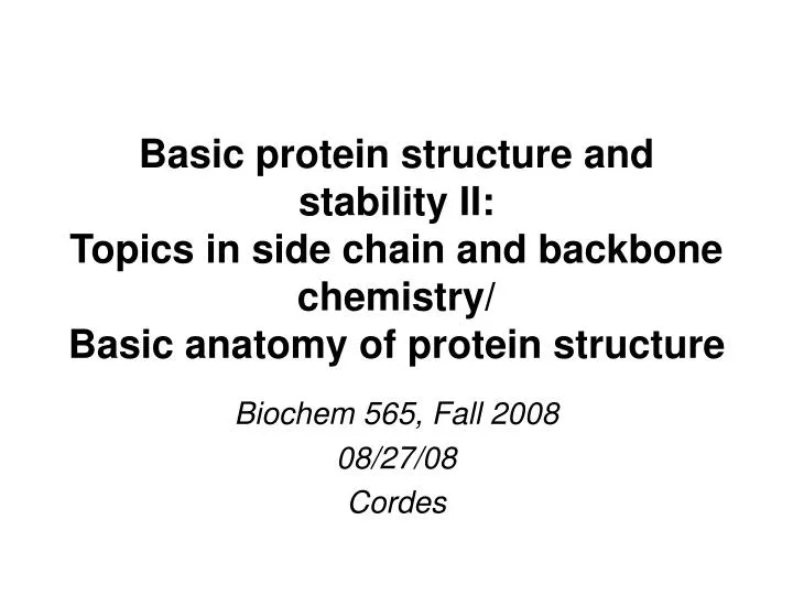
Basic protein structure and stability II: Topics in side chain and backbone chemistry/ Basic anatomy of protein structure. Biochem 565, Fall 2008 08/27/08 Cordes. Covalent chemistry of the side chains and the backbone (main chain). reactions involved in enzymatic catalysis

E N D
Basic protein structure and stability II:Topics in side chain and backbone chemistry/Basic anatomy of protein structure Biochem 565, Fall 2008 08/27/08 Cordes
Covalent chemistry of the side chains and the backbone (main chain) • reactions involved in enzymatic catalysis • covalent posttranslational modifications that regulate activity and processing of proteins, and also cause covalent damage • use of covalent side chain chemistry in analysis of proteins (e.g. crosslinking, attachment of labels, fluorophores etc) • Use of side chain and backbone chemistry in in vitro peptide chemistry Both enzyme catalysis and posttranslational modifications will be covered later in the course. Some other important of the chemistry of side chains will also be covered in a class handout. In lecture today, we will cover just a few interesting, modern examples you may not hear about elsewhere.
Chemistry of Cys thiol group has been exploited in native chemical ligation (NCL) of peptides The nucleophilic character of the thiolate anion of Cys can be used to effectively catalyze specific peptide bond formation between two peptides, if a Cys is present at the N-terminal end of one of the peptides, and if the other peptide is labelled with a thioester at its C-terminus. The Cys thiol exchanges with the thioester, followed by S-N acyl transfer to the amine group of the Cys, forming a peptide bond. The limitation of this reaction is that it requires Cys at the junction between the two peptides: you need an N-Cys peptide. Tam et al Peptide Science60,194 (2001); Muralidahran & Muir, Nature Reviews 3, 429 (2006)
translated sequence of a protein Protein splicing N-extein intein C-extein intein N-extein C-extein splicing intein N-extein C-extein mature protein excised intein
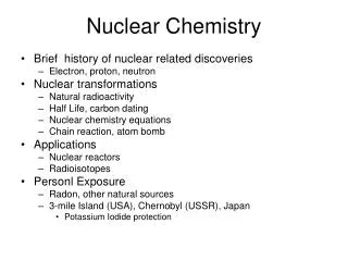
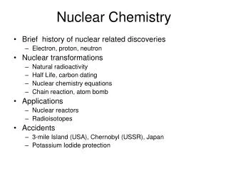
Protein splicing mechanism N-X acyl shift transesterification Asn cyclization X-N acyl shift and succinimide hydrolysis Perler & Adam Curr. Opin, Biotech. 11, 377 (2000)
Protein splicing mechanism at a glance N-X acyl shift transesterification Asn cyclization X-N acyl shift and succinimide hydrolysis Perler & Adam Curr. Opin, Biotech. 11, 377 (2000)
Asparagine deamidation Asparagine deamidation is a major route of protein degradation and damage in vitro and in vivo. It is of concern with regard to the purity and proper function of peptide and protein pharmaceuticals. adapted from Xie L & Schowen, RL J Pharm Sci 88, 8(1999) See also Kossiakoff AA Science 240, 191 (1988)
Oxidative damage/modification of methionine Methionine oxidation is implicated in a variety of aging-related disorders such as cataract formation in the lens and Alzheimer’s disease, though it may also serve nonpathological roles. see for example Kantorow M et al. PNAS101, 9654 (2004; Schoneich Arch Biochem Biophys 397, 302 (2002)
The basic anatomy of protein structure We will now discuss how we look at/describe/analyze protein structures. For Friday, I would like you to read the anatomy section of the web version of JS Richardson’s classic 1981 article the “Anatomy and Taxonomy of Protein Structure”, found at: http://kinemage.biochem.duke.edu/~jsr/index.html This version contains updated notes based on new structural insights since the original article.
The backbone conformation of proteins:combination of regular and irregular structures human hexokinase type I (1QHA) 102 kDa Rosano et al. Structure 1999. pig insulin (1ZNI) 5.7 kDa Bentley et al. Nature 1976. These “ribbon” diagrams show the “skeleton” of a protein. They are a smoothed representation of the “backbone” or “main chain” structure and do not show the side chains
Backbone or main-chain conformation There are three bonds between “main chain” atoms (everything but the side chain) per residue, and torsional rotation can occur about any of these bonds, in principle. Hence, each residue has 3 angles that describe the main chain conformation for that residue.
Backbone conformation:resonance forms of the peptide group The delocalization of the lone pair of the nitrogen onto the carbonyl oxygen shown in the resonance form on the right imparts significant double bond character (40%) to the peptide bond. Breaking of this double bond character by rotation of the peptide bond requires on the order of 18-21 kcal/mol. Consequently there is not free rotation around the peptide bond: rotation about the peptide bond happens on the time scale of seconds/minutes--very slow
Consequences of double bond character in the peptide bond the peptide C-N bond is 0.12A shorter than the Calpha-N bond. and the C=O is 0.02A longer than that of aldehydes and ketones. All six of the atoms highlighted at left lie in the same plane, and as with carbon-carbon double bonds there are two configurations--cis and trans (trans shown at left)
Consequences of double bond character in the peptide bond trans peptide bond cis peptide bond Still another consequence: in the cis form, the R groups in adjacent residues tend to clash. Hence almost all peptide bonds in proteins are in the trans configuration.
...and that means that the dihedral angle describing rotation around the peptide bond, defined by the four atoms Ca(i)-C-N-Ca(i+1), will generally be close to 180°. This angle is known by the greek symbol w. So the properties of the peptide bond place a strong restriction on the backbone conformation or main-chain conformation of proteins, that is to say, the spatial configuration of the non side-chain atoms.
The peptidyl-proline bond cis peptidyl-proline trans peptidyl-proline • The peptidyl proline bond is an exception. • It can be in either the trans or cis configuration, and the equilibrium constant favors the trans only very slightly. • Roughly 20% of all peptidyl proline bonds in native proteins are in the cis configuration. • This is in part because the amide hydrogen is replaced by a methylene group, which can clash with the R group of the preceding amino acid. • Remember that there is still a kinetic effect on the rate of isomerization of the peptidyl proline bond that is similar to that for other peptide bonds--proline cis-trans isomerization can take seconds/minutes to occur, and this can actually limit the rate at which a protein adopts its “native” configuration beginning from a disordered structure (more on this later).
Backbone or main-chain conformation So, one of the three degrees of freedom of the protein backbone is essentially eliminated by the properties of the peptide bond. What about the other two?
f and y angles carboxy (C) terminal end peptide planes Torsional angles are defined by four atoms: y --> Ni-Ca-C’i -Ni+1 f --> C’i-1-Ni-Ca-C’i Notice that these are defined from N to C terminus, using main chain atoms only. A residue’s conformation is usually listed as (f,y), since w is close to 180 for almost all residues. amino (N) terminal end
and between carbonyls Unlike the peptide bond, free rotation occurs about the other two backbone bonds, but steric interactions within the polypeptide still severely limit plausible conformations, so that only certain combinations of phi and psi angles are “allowed” or combinations of an R group, carbonyl or amide group. between R groups 180 not allowed 0 allowed -180 -180 0 180
Steric clashes disallow some f and y combinations Theoretical calculations using hard sphere approximations suggest which phi and psi combinations cause clashes, and between which atoms. Cross-hatched regions are “allowed” for all residue types. The larger regions in the four corners are allowed for glycine because it lacks a side chain, so that no steric clashes involving the beta carbon are possible. from web version of JS Richardson’s “Anatomy and Taxonomy of Protein Structure” http://kinemage.biochem.duke.edu/~jsr/index.html
Observed f and y combinationsin proteins Phi-psi combinations actually observed in proteins with known high-quality structures. Gly residues are excluded from this plot, as are Pro residues and residues which precede Pro (more on this later). Contours enclose 98% and 99.95% of the data respectively. Notice that the observed conformations do not exactly coincide with the theoretically allowed/disallowed confs based on steric clashes. from Lovell SC et al. Proteins 50, 437 (2003)
red= allowed yellow= additionally allowed pale yellow= generously allowed white= disallowed Sample “Ramachandran plot” for a protein The squares denote non-glycine residues, while the triangles are glycines. Glycines have no side chain and are not as restricted because of the lack of side chain steric clashes. This protein has 219 residues, 90% of which are in the “allowed” region and 10% of which are in “additionally allowed” regions. None are in the other regions (except glycines, which don’t count)