Download
1 / 35
350 likes | 585 Views
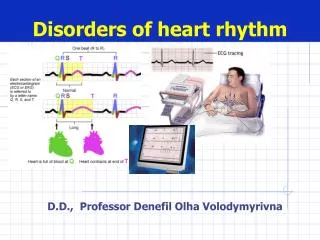
The Electrical Management of Cardiac Rhythm Disorders Bradycardia Device Course. Pacemaker Components. Leads. Epicardial Endocardial. Goals of Cardiac Pacing. The electrical management of bradyarrhythmias requires

E N D
The Electrical Management of Cardiac Rhythm DisordersBradycardiaDevice Course
Leads • Epicardial • Endocardial
Goals of Cardiac Pacing • The electrical management of bradyarrhythmias requires • Ability to deliver enough energy to consistently depolarize the heart (capture) • Ability to correctly sense intrinsic cardiac activity • These functions are affected by many factors • Settings of output parameters (pulse amplitude, pulse width) • Sensitivity parameter settings • Impedance • Electrical concepts
Capture • The capture threshold is defined as the minimum amount of electrical energy required to consistently depolarize the myocardium • When a pacemaker output causes a depolarization, that is also called “capture” • The capture threshold is also called the pacing threshold or the stimulation threshold • The capture threshold is not constant • It can change over time (disease, medications, age) • It can even change over the course of the day!
Capture Threshold Values Threshold values at implant should be low; expect chronic thresholds to increase These are suggested values and may not be possible for all patients
Sensing • Sensing refers to how well the pacemaker is able to “listen to” or perceive intrinsic cardiac events • Important things to consider when talking about sensing • Surface ECG • Intracardiac electrogram (EGM) • Sensing threshold • Sense amplifier • Sensitivity setting and sensitivity safety margin • Unipolar/bipolar configurations • Electromagnet interference
Surface ECG/Intracardiac EGM • Surface ECG: graphic depiction of heart’s electrical signals recorded from electrodes on the body’s surface • Intracardiac EGM: graphic depiction of the heart’s electrical signals recorded by electrodes from inside the heart (pacing lead)
Sensing Threshold = Safety margin Sensitivity Setting Sensitivity Safety Margin • Sensing thresholds are not constant and vary with many factors • The sensitivity safety margin allows reliable sensing even with fluctuations in the sensing threshold • Using this formula, the safety margin should be at least 2 at implant
Electromagnetic Interference (EMI) • EMI is defined as electrical signals of nonphysiologic origin • May interfere with pacemaker (temporarily or permanently) • Common sources of EMI • Cardioversion/defibrillation • Electrocautery • MRIs • Extracorporeal shock wave lithotripsy (ESWL) • Therapeutic radiation • Radiofrequency ablation
What About Cardioversion/Defibrillation? • May permanently damage the pulse generator • Can temporarily inhibit or reprogram the pacemaker • Backup or noise reversion mode • Myocardial thermal damage secondary to shock which may result in ventricular fibrillation, myocardial infarction, or both • Guidelines • Evaluate potential device interactions • Place paddles 4 to 6 inches away from implanted pacemaker • Orient paddles in anterior/posterior position, if possible
What About Electrocautery? • May reprogram or permanently damage the pacemaker • May inhibit the pacemaker • May cause the device to go into backup or noise reversion mode • Myocardial thermal damage secondary to the transmission of the electrical energy may result in VF, MI, or both • Guidelines • Contraindicated
What About MRI? • The magnet in the MRI device can cause asynchronous pacing (pacing without sensing) • Guidelines • Generally contraindicated • Magna-Safe Study
What About Lithotripsy? • The vibrations in extracorporeal shock wave lithotripsy can damage the pacemaker (especially pacemakers with sensors, i.e. rate-adaptive units) • Guidelines • Program to VVI or VOO mode • Keep focal point of lithotripter at least 6 inches away from the implanted pacemaker • Monitor the heart throughout the procedure
What About Therapeutic Radiation? • Damage depends on dose • Damage is cumulative; monitor device throughout course of radiation therapy • Transistors may fail • Pacemakers may fail but mode of failure cannot be predicted • Guidelines • Therapeutic ionizing radiation is contraindicated • If therapeutic radiation is used, pacemaker should be shielded or moved to a less vulnerable location
What About Radiofrequency Ablation? • RF ablation can temporarily or permanently reprogram the pulse generator • Guidelines • Interrogate the pacemaker following the procedure to verify proper function • If necessary, reprogram
Myopotentials • Myopotentials are muscle noises that are sensed by the pacemaker • Can inhibit pacing • The pacemaker senses the myopotential and inhibits the output, thinking the heart has beat on its own! • Can interfere with sensing • Can cause inappropriate pacing • The pacemaker senses myopotential noise and inappropriately “thinks” it is atrial activity; it then tries to pace the ventricle to keep up or track that atrial activity
More EMI Sources • Arc welding • Automobile alternators • Cell phones • Phone antenna should not overlap area of implanted pacemaker • Talk on other side from implanted device • Do not carry an activated cell phone near the implanted pacemaker • May cause inappropriate inhibition, asynchronous pacing, backup mode, inappropriate rate adaptation, and mode switching • Cellular Tested only from St. Jude Medical
EMI in the Medical Environment • Electrocoagulation from electrocautery • Defibrillation • Electroconvulsive therapy • Diathermy • MRI • Stimulators (e.g. transcutaneous nerve) • Dental equipment • Diagnostic ultrasound • Low-frequency acupuncture • Lithotripsy
EMI in the Industrial Environment • Arc welding • Power lines • Transformers • Radio and TV transmitters • Static charge • Large metal frames in magnetic fields • Induction furnaces and heaters • Electrical switches
EMI in the Public Environment • CB radio • Radiofrequency transmissions • Telecommunications antennas • Airport metal detectors • Anti-theft detectors in stores • These may not be marked! • Digital cell phones
Effects of EMI • Pacemaker protection • Hardware backup circuits (to protect against loss of memory or software errors) • Shields • Effects • EMI inhibition: pulse-to-pulse interval extends to the point that the pacemaker does not pace as often as it should. • Noise reversion: change in mode (typically to asynchronous pacing at the programmed rate) which may require reprogramming. • EMI tracking: acceleration of pacing as the pacemaker tries to track electromagnetic signals (“thinking” they are atrial signals)
Pacemaker Overview NASPE / BPEG (NBG) Pacemaker Code
NAPSE/BPEG Generic (NBG) Code Position I II III IV V Category Rate modulation Multisite Pacing Chamber(s) Paced Chamber(s) Sensed Response to Sensing O-None R-Rate modulation Letters Used • O-None • A-Atrium • V-Ventricle • D-Dual • (A+V) • O-None • A-Atrium • V-Ventricle • D-Dual • (A+V) • O-None • T-Triggered • I-Inhibited • D-Dual • (T+I) • O-None • A-Atrium • V-Ventricle • D-Dual • (A+V) S- Single (A or V) S- Single (A or V) Manufacturer’s Designation Only
Magnet Use • Pacemakers • Pace Asynchronously (VOO or DOO) at the given battery rate (Temporarily) • Device will revert back to exactly the same parameters it was programmed to once the magnet is removed • ICD • Will disable ICD Shock Therapy (Temporarily) • Does not affect pacing • Device will revert back to exactly the same parameters it was programmed to once the magnet is removed. • Magnet must be placed over the device in order for temporary changes to occur.
A Systematic Approach to Diagnosing Rhythm Strips • Measure Base Rate • Measure AV/PV Interval • Verify Atrial capture • Verify Atrial sensing • Verify Ventricular capture • Verify Ventricular sensing • Verify Underlying rhythm • Document
Dual Chamber ECG Analysis What is the Analysis? Base Rate 60 ppm MTR 120 ppm AVD 200 ms PVARP 250 ms ECG # 1
Dual Chamber ECG Analysis Base Rate 60 ppm MTR 120 ppm AVD 200 ms PVARP 250 ms What is the analysis? ECG # 2
Dual Chamber ECG Analysis • What is the analysis Base Rate 60 ppm MTR 120 ppm AV 200 ms PV 200 ms PVARP 250 ms ECG # 3
Dual Chamber ECG Analysis • What is the analysis? Base Rate 60 ppm MTR 120 ppm AV 200 ms PV 200 ms PVARP 250 ms ECG # 4
ECG Tracing Results!!! • #1- Normal ECG –Dual chamber pacing and Atrial pacing w/ Ventricular (intrinsic) sensing. • #2- Loss of atrial capture. • #3- Normal ECG • #4-No ventricular sensing and loss of ventricular capture.
Answer • Slide #5 • Normal Sinus Rhythm • Can not determine any pacemaker function • Pacers are usually set to pace above 50 or 60 bpm • Single Chamber ICD- Pace above 40bpm • Pacemakers only work when? • Native heart rate goes below the base rate • An intrinsic beat does not occur before the set Paced and Sensed AV Delays. • Set at an Asynchronous Mode (VOO or DOO)