Download
1 / 63
650 likes | 953 Views
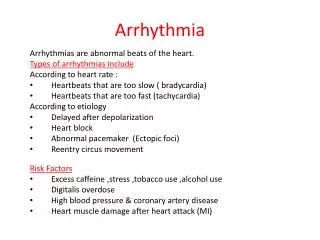
What to do if called for an arrhythmia. Jordan M. Prutkin, MD, MHS Assistant Professor Department of Cardiology/Electrophysiology University of Washington 7/24/2014. What to do…. Check the patient’s pulse Get an ECG Unless there’s no pulse. Then call a code and do ACLS.

E N D
What to do if called for an arrhythmia Jordan M. Prutkin, MD, MHS Assistant Professor Department of Cardiology/Electrophysiology University of Washington 7/24/2014
What to do… • Check the patient’s pulse • Get an ECG • Unless there’s no pulse. • Then call a code and do ACLS
Approaching an EKG • Eyeball • Rate • Rhythm • Axis • Intervals • P waves • QRS • ST-T waves • Overall appearance
Approach to Arrhythmias Do you have calipers? Are there P waves? Are the P waves and QRS’s regular? Are there more P waves than QRS complexes? Are there more QRS complexes than P waves? Is there a constant relationship between the P waves and QRS complexes (constant PR)? Do the QRS complexes look like the baseline QRS (if known)? Are they wider? Narrower?
Case 1 • 73 year old female admitted with pneumonia, reports acute onset of shortness of breath
What does this EKG show? • Sinus rhythm • Atrial fibrillation • Atrial flutter • Atrial tachycardia
What does this EKG show? • Sinus rhythm • Atrial fibrillation • Atrial flutter • Atrial tachycardia
Case 2 • 61 year old male presents to the ED with palpitations • HR 155bpm, BP 122/76
What does this EKG show? • Sinus tachycardia • Atrial fibrillation • Atrial flutter • Atrial tachycardia • Artifact
What does this EKG show? • Sinus tachycardia • Atrial fibrillation • Atrial flutter • Atrial tachycardia • Artifact
Management of Afib/flutter • Is the patient hemodynamically stable? • If there’s hypotension, acute heart failure, mental status change, ischemia, or angina, then cardiovert
If stable, then what? • About 1/2 to 2/3 will terminate spontaneously within 24 hours • Do you need to do anything then? • If rapid or mildly/moderately symptomatic, yes. • Asymptomatic, HR <110bpm • Otherwise, maybe not.
Rate control • IV • Diltiazem 5-20mg IV, then 5-20mg/hr • Metoprolol 5mg IV Q5min x 3 • Esmolol gtt, if in ICU • PO • Diltiazem 30-60mg Q6H • Diltiazem CD 120-240mg Q24H • Verapamil 120-240mg Q24H • Metoprolol 25mg Q6-8H • Metoprolol XL 25-50mg Q12-24H • Atenolol 12.5-50mg Q24H • Digoxin?
Rhythm Control • Amiodarone 150mg IV, then 0.5-1 mg/min gtt • Should really have a central line • Don’t use if afib >48 hours and no anticoagulation • Flecainide • Propafenone Call cardiology • Ibutilide
Anticoagulation/DCCV for AF • Increased risk of stroke after DCCV • If >48 hours, need three weeks of weekly therapeutic coumadin levels, or TEE first • If >48 hours and acute DCCV, give heparin bolus, then infusion and anticoagulate for 4 weeks • If <48 hours, don’t need anticoagulation necessarily • LMWH, dabigatran, rivaroxaban, apixiban okay
Sinus tachycardia • 78 year old admitted with pyelonephritis • HR 120bpm • ECG shows sinus tachycardia
Causes of sinus tach • Fever • Infection/Sepsis • Volume depletion • Hypotension/shock • Anemia • Anxiety • Pulmonary embolism • MI • Heart failure • COPD • Hypoxia • Hyperthyroid • Pheochromocytoma • Stimulants/Illicit substances
Treatment for Sinus Tach • In general, don’t treat heart rate • Treat underlying cause • Exception for acute MI, use beta-blockers
Case 3 • 63 year old male is admitted with chest pain to 5NE • While waiting for a stress test, he reports abrupt onset of palpitations and mild chest discomfort to his nurse. • Pulse 150, blood pressure 132/88
What do you do? • Cry? • Call your senior resident/fellow? • Give metoprolol? • Give adenosine? • All of the above?
What do you do? • Cry? • Call your senior resident/fellow? • Give metoprolol? • Give adenosine? • All of the above?
SVT treatment • Vagal maneuvers (with ECG) • Adenosine (with ECG) • 6mg, 12mg, central line if possible • Beta-blockers/Ca channel blockers (on telemetry) • Can use even if WPW known on baseline ECG • Amiodarone (on telemetry) • Procainamide (on telemetry) • DCCV
What is the diagnosis? • Artifact • Atrial flutter • Atrial tachycardia • Ventricular tachycardia
What is the diagnosis? • Artifact • Atrial flutter • Atrial tachycardia • Ventricular tachycardia
Case 5 • 35 year old male with a history of nonischemic cardiomyopathy • Presents with palpitations
What is the diagnosis? • Atrial fibrillation • Atrial flutter • Sinus Tachycardia • Ventricular Tachycardia
What is the diagnosis? • Atrial fibrillation • Atrial flutter • Sinus Tachycardia • Ventricular Tachycardia
What to do? • If hemodynamically unstable, ACLS/shock • If hemodynamically stable, don’t shock • Call cardiology • Amiodarone 150mg IV, then 0.5-1.0mg/min gtt • Lidocaine 100mg IV, 1-4mg/min gtt • Beta-blocker • IABP • Intubate/paralyze
What to do? • Most times, nothing if asymptomatic • Beta-blocker first line if symptomatic • Check labs? • Usually normal • Turn off telemetry? • Reasonable
Polymorphic VT • Shock/ACLS • Magnesium • Get an ECG when not in VT • Call cardiology • Beta-blocker • Isoproterenol • Pacing • Ischemia evaluation • Avoid QT prolonging drugs (www.torsades.org)
VF • Shock • Do chest compressions • ACLS drugs • Don’t bother with an ECG