Download
1 / 13
130 likes | 281 Views
EMBO Practical Course: Quantitative FRET, FRAP, and FCS Group 1 (Joana Ferreira, Andreas Diepold), Experiment 1a. Single-Pair FRET with TIRF / ALEX setup. Experimental Setup. Calibration of the split camera with fluorescent beads Streptavidin-coating of HF cleaned (!) coverslip

E N D
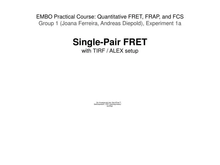
EMBO Practical Course: Quantitative FRET, FRAP, and FCS Group 1 (Joana Ferreira, Andreas Diepold), Experiment 1a Single-Pair FRET with TIRF / ALEX setup
Experimental Setup • Calibration of the split camera with fluorescent beads • Streptavidin-coating of HF cleaned (!) coverslip • Attachment of biotinylated sample
Experimental Setup • Calibration of the split camera with fluorescent beads • Streptavidin-coating of HF cleaned (!) coverslip • Attachment of biotinylated sample: • High FRET control:- Biotinylated double-fluorescently tagged dsDNA
Experimental Setup • Calibration of the split camera with fluorescent beads • Streptavidin-coating of HF cleaned (!) coverslip • Attachment of biotinylated sample: • High FRET control:- Biotinylated double-fluorescently tagged dsDNA • CAP stabilization of transient DNA interaction:- Biotinylated green-labeled half-site DNA 1- Red-labeled half-site DNA 1- cAMP- +/- CAP
Experimental Setup • Calibration of the split camera with fluorescent beads • Streptavidin-coating of HF cleaned (!) coverslip • Attachment of biotinylated sample: • High FRET control:- Biotinylated double-fluorescently tagged dsDNA • CAP stabilization of transient DNA interaction:- Biotinylated green-labeled half-site DNA 1- Red-labeled half-site DNA 1- cAMP- +/- CAP • Data analysis
Experimental Setup • Camera calibration: • Fluorescent beads(green channel, bleeding into red channel) • Manual assignment of corresponding spots Transformation matrix (for the complete experiment) • Data acquisition: • Focusing on spots • Series of pictures(1 sec, 20 fps, alternating green/red excitation) • Data analysis: • Rotation of pictures • Determination of suitable thresholds for the three possible events: Dex/Dem, Dex/Aem, Aex/Aem; choice of events of interest • Automatic analysis of all pictures and matchmaking of corresponding green and red spots • Scatter plot
Results • Positive control – High-FRET DNA FRET efficiency: 0.59 - FWHM: 0.1825
Results • Transcription Factor Detection
Results • Negative control: • Half side 1 – no red signal detection • Add half side 2 – transient binding
Results • CAP • Increase on the red signal, coicindent with green signal • No FRET signal (due to large distance donor - acceptor)
Conclusions Biological: CAP stabilizes the interaction of the two halfs of the CAP binding domains • Technical: • Most useful for immobilized samples (low background noise from mobile molecules). • Single molecule technique Distinction between subpopulations (temporal and spatial resolution) • Distinction between common movement and direct interaction(method works for non-FRETing samples) • Stoichiometry information • Long measurement periods (up to 30 minutes)
Thanks to the tutors! Robert Crawford Alex Kiel Kostas Lymperopoulos Achillefs Kapanidis Dirk-Peter Herten
Thanks for your attention! Andreas Diepold Joana Ferreira