Download
1 / 6
60 likes | 270 Views
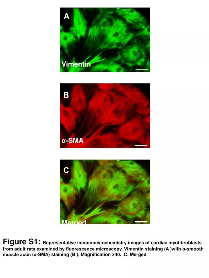
A. Vimentin. B. α -SMA. C. Merged. Figure S1: Representative immunocytochemistry images of cardiac myofibroblasts from adult rats examined by fluorescence microscopy. Vimentin staining (A )with α-smooth muscle actin (α-SMA) staining (B ). Magnification x40. C: Merged. Col III.

E N D
A Vimentin B α-SMA C Merged Figure S1: Representative immunocytochemistry images of cardiac myofibroblasts from adult rats examined by fluorescence microscopy. Vimentin staining (A )with α-smooth muscle actin (α-SMA) staining (B ). Magnification x40. C: Merged
Col III α-Tubulin Figure S2: Cardiac protein levels of collagen III (col III). Quantification of band intensities was measured by densitometry and normalized to respective α-tubulin. Values are mean±SEM of 6 animals. *p<0.05.
Figure S3: Effect of different doses of leptin on the proliferation in cardiac myofibroblasts at 24 hours. Cell proliferation was determined by an MTT assay. Data are expressed as percent of controls (vehicle, v). Values are mean±SEM of four assays. 120 % (vs CT) 60 0 V 10 50 100 Leptin (ng/ml)
V Leptin Leptin Leptin + Mel + Rap 1.5 1 Activity MMP-2 0.5 0 V Leptin Leptin Leptin + Mel + Rap Figure S4: The effect of cells stimulated with leptin (100ng/ml) in presence or absence of melatonin (10-3 mmol/L) and rapamycin (10-4 mmol/L) on metalloproteinase (MMP) 2 activity in cardiac myofibroblasts
Vehicle 150 Leptin * Leptin + Rap † * Figure S5: Superoxide anion production in cells stimulated with leptin (100 ng/ml) at different times in presence or absence of rapamycin (10-4 mmol/L).Values are mean±SEM of four assays. *p<0.001 vs vehicle (v, control conditions); † p<0.001 vs leptin. * * † † 100 * * Fluorescence (A.U.) 50 0 V 1 2 4 (hours)
LEPTIN Cardiac myofibroblasts mTOR activation ROS MMP-2 activity TGF-β Galectin-3 CTGF Degradation Collagen I Synthesis Collagen I Figure S6: Scheme illustrating the possible mechanism involved in the fibrotic effect induced by leptin in cardiac myofibroblasts.. Leptin could favour collagen deposition by modulating both its synthesis and also its degradation. Leptin induced collagen synthesis by increasing ROS (reactive oxygen species) production directly or through the activation of mTOR pathway. Galectin-3 and growth factors such as TGF-β and CTGF seem to be the final mediators of the collagen synthesis induced by leptin. The reduction in MMP-2 (metalloproteinase-2) activity induced by leptin seems to involve an oxidative stress- and mTOR pathway-independent mechanism. EXTRACELLULAR MATRIX