Download
1 / 5
80 likes | 582 Views
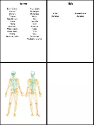
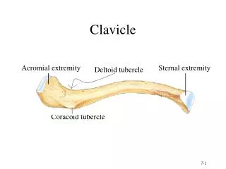
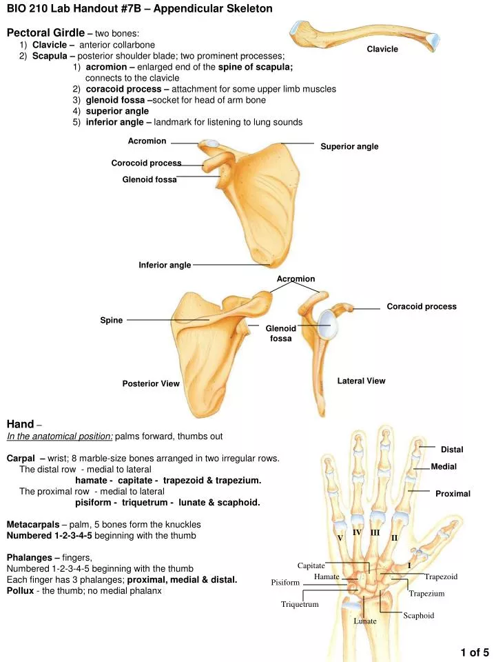
BIO 210 Lab Handout #7B – Appendicular Skeleton Pectoral Girdle – two bones: 1) Clavicle – anterior collarbone 2) Scapula – posterior shoulder blade; two prominent processes; 1) acromion – enlarged end of the spine of scapula; connects to the clavicle

E N D
BIO 210 Lab Handout #7B – Appendicular Skeleton Pectoral Girdle – two bones: 1) Clavicle – anterior collarbone 2) Scapula – posteriorshoulder blade; two prominent processes; 1) acromion – enlarged end of the spine of scapula; connects to the clavicle 2) coracoid process – attachment for some upper limb muscles 3) glenoid fossa –socket for head of arm bone 4) superior angle 5) inferior angle – landmark for listening to lung sounds Clavicle Acromion Superior angle Corocoid process Glenoid fossa Inferior angle Acromion Coracoid process Spine Glenoid fossa Posterior View Lateral View Hand – In the anatomical position: palms forward, thumbs out Carpal – wrist; 8 marble-size bones arranged in two irregular rows. The distal row - medial to lateral hamate - capitate - trapezoid & trapezium. The proximal row - medial to lateral pisiform - triquetrum - lunate & scaphoid. Metacarpals – palm, 5 bones form the knuckles Numbered 1-2-3-4-5 beginning with the thumb Phalanges – fingers, Numbered 1-2-3-4-5 beginning with the thumb Each finger has 3 phalanges; proximal, medial & distal. Pollux - the thumb; no medial phalanx Distal Medial Proximal IV III II V Capitate I Hamate Trapezoid Pisiform Trapezium Triquetrum Scaphoid Lunate 1 of 5
Greater tubercle Lesser tubercle Deltoid tuberosity Coronoid fossa Olecranon fossa Lateral epicondyle Capitulum Medial epicondyle Trochlea Posterior view Upper Limb Humerus – brachium; articulates with the scapula and with the radius & ulna at the elbow 1) head – smooth & hemispherical, proximal end and fits into the glenoid fossa of the scapula 2) greater tubercle – lateral 3) lesser tubercle – medial 4) deltoid tuberosity – midway on the lateral side, v-shaped and rough; deltoid muscle attachment 5) capitulum – dital & lateral ball-like condyle: articulates with radius 6) trochlea – medial condyle, hourglass tipped on its side; articulates with ulna 7) coronoid fossa – superior to the trochlea on anterior surface; 8) olecranon fossa – posterior surface, superior to trochlea 9) medial “funny bone” & lateral epicondyles – muscle attachment points For anterior view, Place Head in superior position Radial & coronoid fossas are visible. Larger, deeper olecranon fossa is posterior Right forearm in anatomical position Antebrachium - 2 parallel bones Rradius – carries the hand; lateral bone in anatomical position; Head – proximal, disc-shaped, articulates with humerus capitulum Radial tuberosity – prominence on medial aspect of shaft; tendon of biceps muscle attaches here styloid process – attachment for wrist ligaments Ulna – forms the elbow joint with the humerus; medial bone of the forearm Olecranon process – grips the trochlea to form a hinge joint; Coronoid process – grips the trochlea to form elbow hinge joint; Trochlear notch – deep concavity between olecranon & coronoid processes Styloid process – ligament runs to the wrist Head Radial tuberosity Ulna Radius Olecranon process Styloid process Styloid process Trochlear notch Coronoid process 2 of 5 Ulna
Pelvic Girdle – 2os coxae (hip bones) united anteriorly at the pubic symphysis and posteriorly at the sacrum and coccyx Os coxae consist of 3 separate bones in childhood - ilium, ischium & pubis. In adults, these bones are firmly fused. Names are retained to refer to different regions. Acetabulum - deep socket where these 3 bones fuse; receives the head of the femur. Ilium – large flaring region, major part of coxal bone; sacroiliac joint – joint where os coxae are connected posteriorly iliac crest – superior margin, rough, where you rest your hands on your hips anterior superior iliac spine – where the iliac crest terminates anterior inferior iliac spine Ischium – the “sit-down bone” the most inferior and posterior position of the coxal bone ischial tuberosity – most outstanding marking of the ishium; receives the weight of the body when sitting Ischial spine – projects medially into the pelvic cavity, point of ligament attachment greater sciatic notch – large notch superior to ischial spine; sciatic nerve and blood vessels pass through here lesser sciatic notch – small notch just inferior to the ischial spine Pubis – most anterior of the coxal bone; V-shaped with two rami; in the anatomical position it is horizontal and the bladder rests upon it pubic symphysis – fibrocartilage disc; formed midline where two pubic bones are joined Obturator foramen – the 2 rami of the pubis run laterally to join Ischium; a few blood vessels & nerves pass through nearly closed by a fibrous membrane; obturator = closed Iliac crest Ilium Sacroilliac joint Ilium Anterior superior iliac spine Sacrum Anterior inferior iliac spine Greater sciatic notch Acetabulum Ischial spine Obturator foramen Pubis Lesser sciatic notch Ischium Ischial tuberosity Pubic symphysis 3 of 5
Femur – thigh bone; heaviest, strongest bone in the body; Head – ball-like; articulates with the hip bone in deep acetabulum Fovea capitis femoris – “pit of the head”; ligament attachment securing head to acetabulum Neck – carries the head; angles laterally to join the shaft; broken hips Greater trochanter – superior position where the neck joins the shaft; Lesser trochanter – inferior position posterior and medial Linea aspera – “rough line” a long vertical ridge that appears to diverge distally Medial condyle – distal end; wheel-like; same side as femur head; articulates with tibia Lateral condyle – distal end; opposite femur head; articulates with leg tibia Patella – knee cap; within a tendon that secures thigh muscles to tibia Apex – pointed end where tendon attaches Base – flattened end; Greater trochanter Neck Head Lesser trochanter Base Fovea capitis femoris Linea aspera Patella Apex Medial condyle Lateral condyle Leg – two bones 1) Tibia – “shin bone” is larger, more medial; articulates with femur to form knee Tibial tuberosity – inferior to the condyles of proximal end; rough; ligament attachment Intercondylar eminence (tubercles) – irregular projections between condyles Anterior crest – shaft is triangular in cross section; a sharp ridge of bone on the anterior surface; Medial malleolus – “little hammer”; rounded bony process at distal tip of tibia; medial bulge of the ankle 2) Fibula– lateralstick-like bone; shaft is heavily ridged for muscle attachment; articulates with tibia Lateral malleolus – distal end of fibula; forms conspicuous lateral ankle bulge; articulates with the talus Anterior crest – same as tibia Patella Lateral condyle Medial condyle Fibula Anterior crest Anterior crest Tibia Medial malleolus Lateral malleolus 4 of 5
Calcaneus Talus Cuboid Navicular Lateral cuneiform Intermediate cuneiform V Medial cuneiform IV II Metatarsals I Proximal Phalanges Medial Distal Intermediate cuneiform #5 Medial cuneiform #4 #2 #1 Digits Foot – tendons of foot muscles hold bones firmly in domed position to form 3 strong arches; Tarsals – corresponds to carpals of hand; 7 bones; body weight is carried by 2 largest most posterior bones 1) talus – “ankle” articulates with tibia & fibula superiorly 2) calcaneus – “heel bone”; Achilles tendon attaches here 3) navicular – articulates with the talus and anteriorly with cuneiforms 4,5,6) medial cuneiform, intermediate cuneiform, lateral cuneiform form the most anterior row; articulate with metatarsals 1,2 & 3 respectively 7) cuboid – articulates with the calcaneous; anteriorly with metatarsals 4&5 Metatarsals – corresponds to metacarpals of hand; 5 bones numbered 1-2-3-4-5; always begin with the big toe. The enlarged head of metatarsal #1 forms the ball of the foot Phalanges - 14 bones; smaller than the fingers but general structure is same; 3 phalanges to each toe - proximal, medial & distal Hallux – big toe; only has 2 phalanges, proximal and distal III Medial #3 5 of 5