Download
1 / 27
480 likes | 1.36k Views
Tissues of the Periodontium. Presented by: Rita Ann Classe , RDH, BS. What’s foundation got to do with it?. The Periodontium. What is it? Functional system of tissues that surrounds the teeth and attaches them to the jawbone. Why is it important?

E N D
Tissues of the Periodontium Presented by: Rita Ann Classe, RDH, BS
The Periodontium • What is it? • Functional system of tissues that surrounds the teeth and attaches them to the jawbone • Why is it important? • Essential to the understanding of the normal function of the periodontium, disease prenvention, and the periodontal disease process
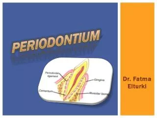
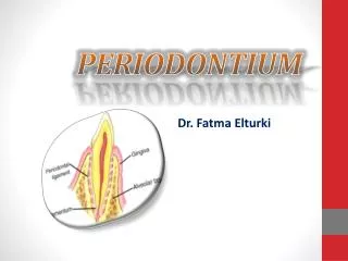
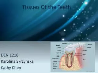
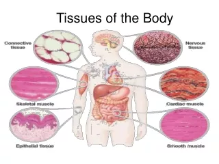
Tissues of the Periodontium Gingiva Cementum Periodontal ligament Alveolar bone
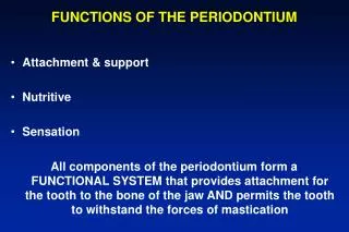
Functions of the Periodontium • Provides seal around cervical portion (neck) of tooth • Holds tissue against tooth during mastication • Suspends and maintains tooth in socket • Protects underlying dentin • Anchors the ends of the periodontal ligament fibers to the tooth • Surrounds and supports the roots of the tooth
Gingiva • Provides a tissue seal around the cervical portions of the teeth and the alveolar processes of the jaw
Gingiva • Boundaries: • Gingival margin- coronal boundary • Alveolar mucosa- apical boundary • Demarcations: • Free gingival groove- separates free and attached gingiva • Mucogingival junction- where attached gingiva meets alveolar mucosa
Functions of Gingiva • Provides seal around cervical portion of tooth • Holds tissue against tooth during mastication
Anatomical Areas of Gingiva • Free gingiva (unattached or marginal) • Attached gingiva • Interdental gingiva • Gingival Sulcus
Free Gingiva • Unattached portion that surrounds tooth in area of cementoenamel junction (CEJ) • Fits closely around tooth but not directly attached (turtleneck or cufflike) • Meets tooth in a thin rounded edge called the gingival margin • Gingival margin follows contours of teeth creating a scalloped outline
Attached Gingiva • Tightly connected to cementum on cervical third of root and to periosteum of alveolar bone • Lies between free gingiva and alveolar mucosa • Pale/coral pink or light brown/black in color • Stippling (orange peel) texture • Withstands mechanical forces • Prevents free gingiva from being pulled away from tooth
Interdental Gingiva • Fills area between 2 adjacent teeth • Facial and lingual • Col=valley-like depression that lies apical to contact area • Prevents food impaction
Gingival Sulcus • V-shaped, shallow space around tooth • Located between the free gingiva and tooth surface • Base of sulcus formed by junctional epithelium • Depth in health is 1-3mm
One function of the gingva is to provide a seal around the cervical portion of the tooth. TRUE FALSE
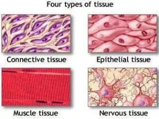
Periodontal Ligament (PDL) • Layer of soft connective tissue that covers root and attaches it to the bone of the tooth socket • Composed mainly of fiber bundles • Fibers of PDL attach on one side to the root cementum and on the other side to the alveolar bone of tooth socket
Functions of PDL • Supportive • Sensory • Nutritive • Formative • Resorptive
Cementum • Layer of hard, mineralized tissue that covers dentin surface of root • Light yellow in color • Resistant to resorption • Receives nutrients from the PDL • No nerve or blood supply
Functions of Cementum • Anchors PDL to tooth • Protects underlying dentin • Compensates for occlusal tooth wear
The function of the PDL is to protect the underlying dentin. TRUE FALSE
Alveolar Bone (Alveolar Process) • Bone of the upper or lower jaw that surrounds and support the roots of the teeth • Dependent on presence of tooth
Function of Alveolar Bone • Forms the bony sockets that provide support and protection for the roots of the teeth
Components of Alveolar Bone • Alveolar bone proper (cribiform plate) • Alveolus • Cortical bone • Alveolar crest • Cancellous bone (spongy bone) • Periosteum
Let’s Review • Provides seal around cervical portion (neck) of tooth • Holds tissue against tooth during mastication • Suspends and maintains tooth in socket • Protects underlying dentin • Anchors the ends of the periodontal ligament fibers to the tooth • Surrounds and supports the roots of the tooth
References Nield-Gehrig, J.S. and Willmann, D.E. (2008). Foundations of Periodontics for the Dental Hygienist, Second Edition. Baltimore, MD: Lippincott Williams & Wilkins.