Download
1 / 77
800 likes | 1.07k Views
Cell Unit III: Cell Division, Cell Cycle, Transcription and Translation. Chapters 12, 13, 16, 17. Limits to Cell Growth. The larger a cell becomes, the more demands a cell places on its DNA If extra copies of DNA are not made, an “information crisis” would occur

E N D
Cell Unit III: Cell Division, Cell Cycle, Transcription and Translation Chapters 12, 13, 16, 17
Limits to Cell Growth • The larger a cell becomes, the more demands a cell places on its DNA • If extra copies of DNA are not made, an “information crisis” would occur • The cell also has more trouble moving nutrients and wastes across the cell membrane • Food, oxygen, water, and wastes move through the cell membrane • The rate at which the exchange takes place depends on the surface area of the cell • The rate at which food and oxygen are used up and wastes produced depends on the cell’s volume
Ratio of Surface Area to Volume • Volume increases much more rapidly than surface area causing the ratio of surface area to volume to decrease • This decrease creates serious problems for the cell such as: • Inability to remove wastes from the cell • Lack of sufficient oxygen and food entering through the cell membrane
Division of the Cell • The process by which a cell divides into two new daughter cells is called cell division • Before cell division occurs, the cell replicates, or copies, all of its DNA • Each daughter cell gets one complete set of genetic information • Each daughter cell also has an increased ratio of surface area to volume
Cell Division • Each cell has only one set of genetic information • must be copied before cell division begins • The first stage, division of the cell nucleus, is called mitosis • The second stage, division of the cytoplasm, is called cytokinesis • Reproduction by mitosis is classified as asexual • Mitosis is the source of new cells when a multicellular organism grows and develops
Chromosomes • Chromosomes are made of DNA (genetic information) and proteins (histones) • The cells of every organism have a specific number of chromosomes • Fruit flies = 8, human = 46, carrots = 18 • Chromosomes are not visible in most cells except during cell division • Each chromosome consists of two identical sister chromatids which separate during cell division • Each pair of chromatids is attached in an area called the centromere
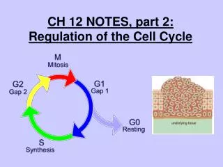
The Cell Cycle • Interphase is the period in between periods of cell division • The cell cycle is the series of events that cells go through as they grow and divide • During the cell cycle, a cell grows, prepares for division, and divides to form two daughter cells, each of which then begins the cycle again • The cell cycle consists of four phases • M, S, G1, and G2
Events of the Cell Cycle • During the normal cell cycle, interphase can be quite long, whereas the process of cell division takes place quickly • The G1 phase is a period in which cells do most of their growing • In the S phase, chromosomes are replicated and the synthesis of DNA molecules takes place • During the G2 phase, many of the organelles and molecules required for cell division are produced
Mitosis • Prophase: • Chromosomes become visible, centrioles begin to organize the spindle and move to opposite ends of the cell, fibers attach to centromeres, nucleolus and nuclear envelope disappear • Metaphase: • Chromosomes line up across the center of the cell • Anaphase: • Centromeres split and individual chromatids are separated into two groups near the poles • Telophase: • Chromosomes disperse, nuclear envelope and nucleolus re-form, spindle breaks apart
Cytokinesis • Cytokinesis is the division of the cytoplasm itself and usually occurs at the same time as telophase • In most animal cells, the cytoplasm is drawn inward until the cytoplasm is pinched into two nearly equal parts. This is called a cleavage furrow. • In plants, a structure known as the cell plate forms midway between the divided nuclei
Controls on Cell Division • When placed on a petri dish with a thin layer of nutrient solution, cells will grow until they form a thin layer on the bottom of the dish • When cells come into contact with other cells, they respond by not growing • If cells are removed from the center of the dish, the cells bordering the open space will divide until they have filled the space • Controls on cell growth and division can be turned off and on
Cell Cycle Regulators • Several scientists discovered that cells in mitosis contained a protein that when injected into a nondividing cell, would cause a mitotic spindle to form • They called this protein cyclin because it seemed to regulate the cell cycle • Cyclins regulate the timing of the cell cycle in eukaryotic cells • Proteins that respond to events inside the cell are called internal regulators • External regulators respond to events outside of the cell
Uncontrolled Cell Growth • Cancer is a disorder in which some of the body’s own cells lose the ability to control growth • Cancer cells do not respond to the signals that regulate the growth of most cells • They divide uncontrollable and form masses of cells called tumors that can damage the surrounding tissues • Causes include smoking, radiation, and viral infections • Damaged or defective p53 genes cause the cells to lose the information needed to respond to signals that would normally control their growth
p53 is a protein that functions to block the cell cycle if the DNA is damaged. If the damage is severe, this protein can cause apoptosis (cell death). • p53 levels are increased in damaged cells. This allows time to repair DNA by blocking the cell cycle. • A p53 mutation is the most frequent mutation leading to cancer. • p27 is a protein that binds to cyclin and cdk blocking entry into S phase. Recent research (Nature Medicine 3, 152 (1997)) suggests that breast cancer prognosis is determined by p27 levels. Reduced levels of p27 predict a poor outcome for breast cancer patients.
CHAPTER 13: MEIOSIS AND SEXUAL CYCLES Meiosis - cell division that reduces the diploid # to the haploid # in the formation of sex cells (gametes). Example (Humans) - 46 chromosomes is reduced to 23. MOST IMPORTANT - the cells produced at the end of meiosis contain one chromosome of each homologous (matching) pair.
TERMS: GENE - HEREDITARY INFORMATION, IN A SECTION OF A DNA MOLECULE ON A CHROMOSOME. LOCUS (LOCI) - A GENE’S SPECIFIC LOCATION ON A CHROMOSOME. CLONE - A GROUP OF GENETICALLY IDENTICAL INDIVIDUALS ( WHAT MITOSIS PRODUCES) ASEXUAL REPRODUCTION - REPRODUCTION W/O SEX (NO MALE/FEMALE; 1 PARENT; OFFSPRING IS A CLONE OF PARENT. HOMOLOGOUS CHROMOSOMES - A MATCHING PAIR ALWAYS ONE FROM EACH PARENT.(one paternal/ one maternal.)
AUTOSOMES - CHROMOSOMES NOT DIRECTLY INVOLVED IN DETERMINING SEX. (IN HUMANS: 22 HOMOLOGOUS PAIR). SEX CHROMOSOMES - THE CHROMOSOMES DIRECTLY INVOLVED IN DETERMINING SEX (IN HUMANS THE LAST HOMOLOGOUS PAIR). (a) CALLED (X) & (Y) CHROMOSOMES. (b) XX = FEMALE & XY = MALE. (c) In other organisms: (1) Insects (Grasshoppers, Roaches): X-O sex chromosomes. O represents no sex chromosome = Male (2) Birds, Butterflies and some fish: Z-W sex chromosomes. Female gamete determines sex. Males are ZZ, Females are ZW (3) Parthenogenesis – wasps, bees and ants. If the egg is fertilized it becomes a female and is diploid. If the egg is unfertilized it is male and haploid. FERTILIZATION (or SYNGAMY) - UNION OF GAMETES. KARYOTYPE: DISPLAY OF AN INDIVIDUAL’S CHROMOSOMES. CHROMOSOMES ARE COLLECTED DURING METAPHASE. THIS IS DONE BY NUMBER, SIZE & TYPE CHROMOSOME.
THE HUMAN LIFE CYCLE: (characteristic of most animals) Gametes are the only haploid cells. The diploid zygote divides by mitosis producing a diploid organism.
MEIOSIS STEPS:( FIG. 13.5.) (a) Each chromosome replicates. (This shows 1 homologous pair). Remember - sister chromatids & centromere. (b) Meiosis I segregates the homologous pair into 2 different cells (each new daughter cell is in HAPLOID). (c ) Meiosis II separates sister chromatids into chromosomes. No chromosome duplication)
MEIOSIS TERMS: Synapsis - ( in prophase I ) - the duplicated chromosomes pair with their Homologues). This is a PROCESS.Homologous chromosomes made of two sister chromatids come together as pairs. Homologue - one of a homologous pair. Tetrad - the four closely associated chromatids of a homologous pair together. This happens during synapsis. Crossing over - (a process) reciprocal exchange of genetic material between nonsisterchromatids.
COMPARING MITOSIS & MEIOSIS. MEIOSIS - Prophase I with -(a) Tetrad & synapsis making a synaptonemal complex (b) Crossing over with the chiasma. MITOSIS- No tetrads, synapsis, or crossing over. DAUGHTER CELL DIFFERENCE - Mitosis has produced 2 identical cells. Meiosis produced daughter cells with one of each homologous pair.
FIG. 13.8- This shows the mostimportant concept of meiosis (how it produces genetic variation in organisms). INDEPENDENT ASSORTMENT: At the end of meiosis chromosome pairs distribute themselves independently of one another. This causes 4 different combinations of chromosomes with 2 homologous pair.
1st MEIOTIC DIVISION RESULTS IN INDEPENDENT ASSORTMENT OF MATERNAL & PATERNAL CHROMOSOMES IN DAUGHTER CELLS. FORMULA: The number of combinations possible when chromosomes assort independently into gametes during meiosis is 2n, where (n) is the haploid # in the organism. EXAMPLE - Human haploid (n) is 23. 223 is over 8 million. A male can produce 8 million genetically different combinations of sperm & a female 8 million combinations of eggs. RANDOM FERTILIZATION then would produce 8 million x 8 million(over 64 Trillion) possibly different genetic combinations in the offspring.
Crossing Over -produces individual chromosomes that combine genes inherited from our two parents. Independent Assortment, Random Fertilization, & Crossing Over - result ways that genetic variation can be produced.
SUMMARY: Prophase I & Anaphase I produce the most variation in the 4 new daughter cells. If clones were genetically different, this would be due to mutation (change in the code of DNA). Remember these !!!! Which might be a daughter cell of meiosis I ? Which might be a daughter cell of meiosis II?
CHAPTER 16 - THE MOLECULAR BASIS OF INHERITANCE DNA - most celebrated molecule of all time. It is made of nucleic acids that have the unique ability to direct their own replication. PROBLEM: Since a chromosome is made of protein & DNA which one is carrying the genetic material? There can be an infinite # of proteins so it would be a prime candidate to carry genetic material.
JAMES WATSON – CO-FOUNDER OF THE STRUCTURE OF DNA Watson & Crick working on the DNA structure model. (April 1953)
Transformation of Bacteria - Frederick Griffin.(1928) The captions under the picture is all that is needed to explain this experiment. TRANSFORMATION - the change in genotype & phenotype due to the assimilation of external DNA by a cell.
EVIDENCE THAT VIRAL DNA CAN PROGRAM CELLS (FIG. 16.2) Virus is made of a protein coat & DNA core. Virus injects DNA into a Bacteriophage. DNA coat has radioactive protein coat (S35) while DNA is radiated with (P32). HERSHEY-CHASE EXPERIMENT That the bacteria are called T2 Phages.
ADDITIONAL EVIDENCE THAT DNA IS THE GENETIC MATERIAL OF CELLS Erwin Chargaff - Said that the bases of DNA (A, T, C, G) vary from one species to another. He also found a regular ratio of bases. (A approximately = T; and G approx. = C). This was known as Chargaff’s Rules. NOTE: All these discoveries were before Watson & Crick discovered the double helix structure of DNA.
Structure of a DNA strand DNA is composed of nucleotides ( 5 carbon sugar, phosphate & a nitrogenous base (A,T,C,G). Phosphate of one nucleotide is attached to the sugar of the next nucleotide.
Fig. 16.5 (a) The Double Helix Structure of DNA. Adenine (A) is always paired with Thymine (T) & Guanine (G) is always paired with Cytosine (C). The nitrogenous bases are held together with Hydrogen bonds (weak). We even know the distances between steps of the DNA rungs. What’s a nm?
Notice the strands are oriented in opposite directions. This entire structure was worked out by Watson & Crick in 1953 with help from Rosalind Franklin’s x- ray diffraction photo of DNA
Base Pairing in DNA. A & G are double ring compounds called Purines. T & C are single ring compounds called Pyrimidines. Each rung of DNA is made of a Purine attached to a Pyrimidine. Held together by H bonds.
Meselson& Stahl tested the three hypothesis's on DNA replication
Beginnings of how DNA Replicates. Elongation of DNA at a replication fork is catalyzed by a enzyme called DNA polymerase. Rate of elongation in humans is approx.50/sec.